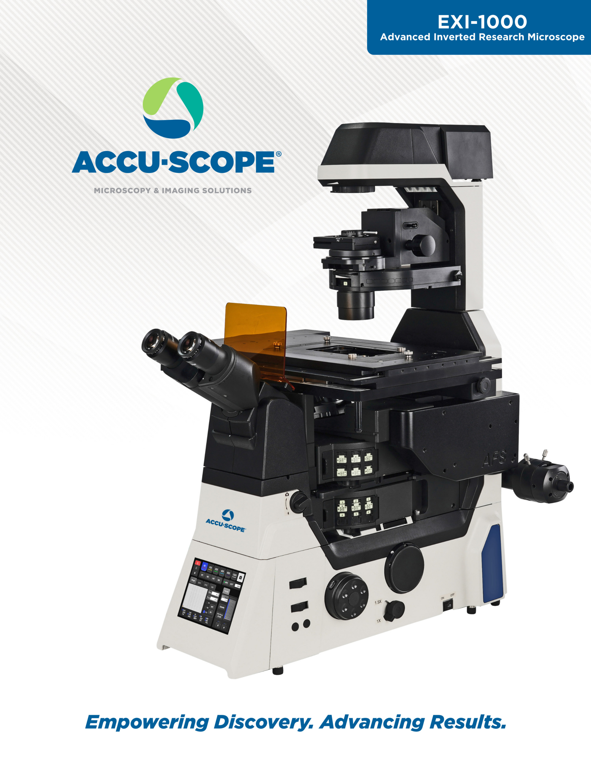


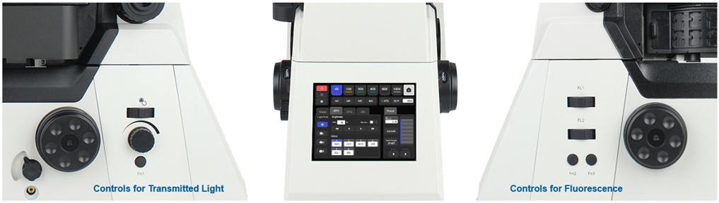
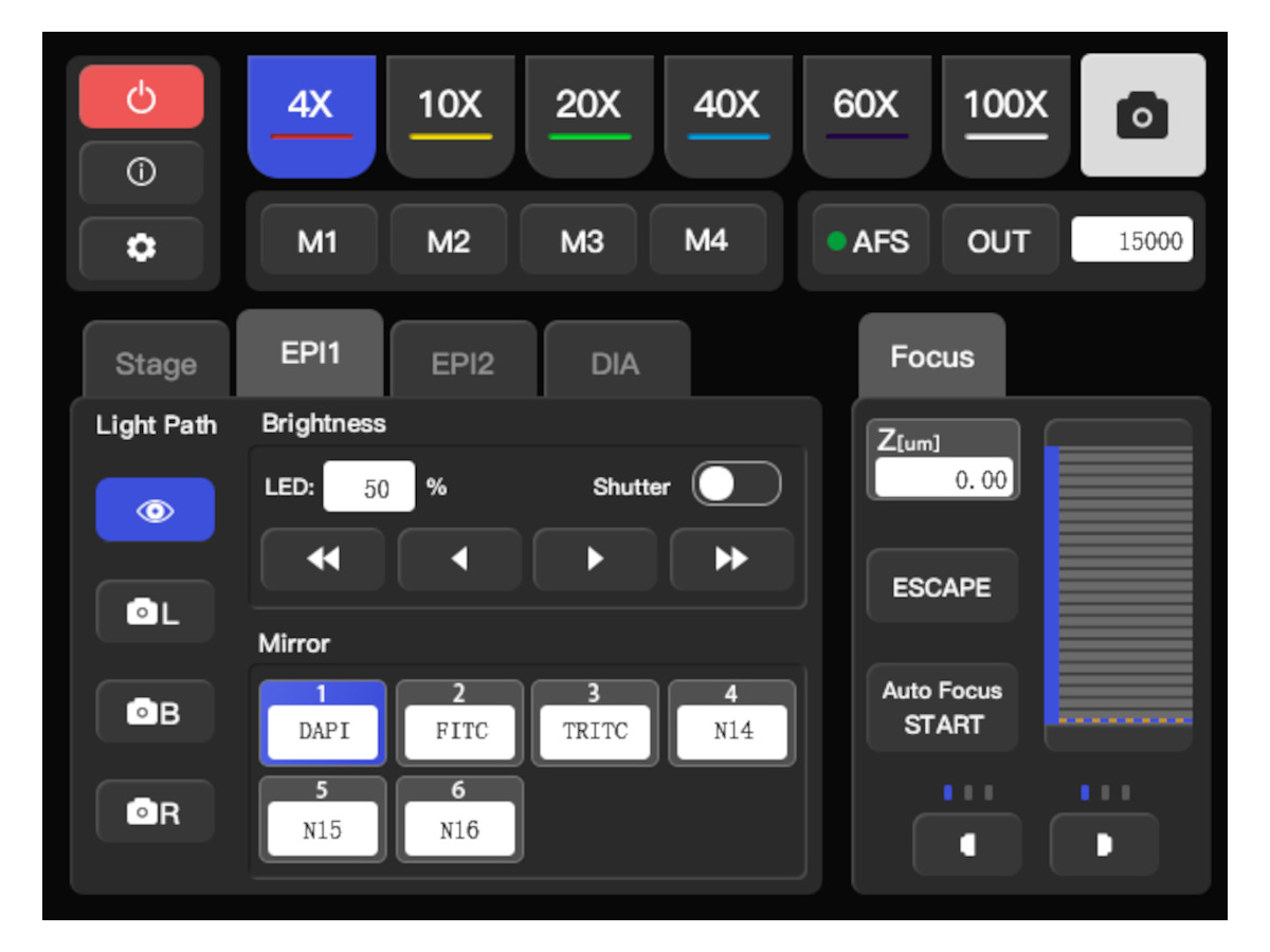
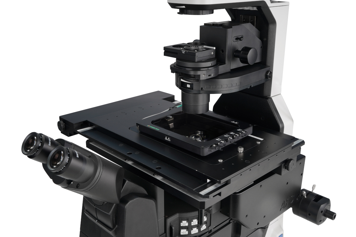
The EXI-1000 puts everything you need to know — and everything you need to control — right at your fingertips. The 5.6-inch front-panel touchscreen displays real-time system status at a glance: current objective magnification, fluorescence channel, illumination intensity, imaging port, Z position, and the state of every motorized component. No hunting through software menus or guessing what your system is doing mid-experiment.
For those moments when you’d rather keep your hands off the screen, dedicated knobs and buttons on both sides of the panel give you tactile, eyes-free control — a small but meaningful detail when you’re working in a darkened lab with a sample under the scope. Motorized components including the objective turret, fluorescence cube, condenser, camera port, and Z-axis are all accessible from the same interface, so switching between modes or adjusting on the fly never interrupts your workflow.
The result is a microscope that stays transparent — giving you instant situational awareness without demanding your attention — so you can stay focused on the experiment, not the instrument.
Live-cell imaging experiments can run for hours or even days — and throughout that time, your cells are sensitive to the world around them. Small fluctuations in temperature, humidity, or CO₂ levels can trigger stress responses, alter cell behavior, or compromise viability, quietly undermining the biological relevance of your data without any obvious sign that something has gone wrong. By the time you realize the results look off, the experiment may be unrecoverable.
Maintaining a stable, physiologically appropriate environment isn’t optional — it’s the difference between data you can publish and data you have to repeat.
The EXI-1000 is designed to accommodate a wide range of stage-top incubators and full microscope enclosures from leading third-party manufacturers. Stage-top incubators fit directly onto the microscope stage, creating a compact, tightly controlled environment around your sample. Microscope enclosures take a broader approach, surrounding the entire upper portion of the instrument to maintain consistent conditions across the stage, objectives, and optical path. Both approaches minimize environmental fluctuations so your cells remain healthy, your biology stays relevant, and your experiment runs to completion — on the first try.
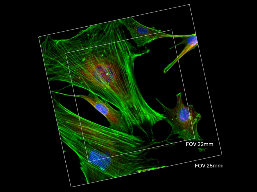
Scientific cameras have advanced dramatically — larger sensors, faster readout, greater sensitivity — and the microscope platform underneath them needs to keep pace. A camera can only deliver on its full potential when the optical system feeding it is wide enough to fill the sensor. That’s where many conventional research microscopes fall short: with a maximum field of view of 22mm, they leave a meaningful portion of a modern large-format sensor unused, sacrificing data and throughput before the experiment even begins.
The EXI-1000 is built for where imaging is today — and where it’s heading. Its 25mm field of view, matching the standard set by top-tier research platforms, ensures that the full imaging area of today’s large-sensor scientific cameras is put to work. The result is more cells in every frame, more events captured in every time point, and more biological context in every image — without any increase in acquisition time. For screening workflows, tiled imaging, or any experiment where throughput and coverage matter, that difference is significant.
Investing in an imaging platform is a long-term decision. The 25mm FOV of the EXI-1000 ensures that as cameras continue to evolve, your microscope won’t be the bottleneck — leaving you free to adopt new technology and push your science forward without being held back by the instrument beneath it.
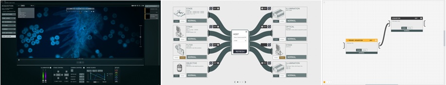
Starlyte Nebula is purpose-built acquisition software that unifies your entire microscopy workflow — from experiment design to image capture — in a single, intuitive interface. Rather than juggling separate driver environments, scripting tools, and acquisition GUIs, researchers can connect their cameras, stages, lasers, filter wheels, and objectives all in one place, with live device status and hot-swap support for over 30 compatible devices. Nebula’s visual, node-based automation engine lets you chain triggers, multi-channel acquisitions, time lapses, Z-stacks, and X/Y or lambda scans into reusable workflows that can be saved once and run indefinitely — so your protocols are reproducible, shareable across the lab, and ready to scale. Whether you’re a researcher who wants to get data out fast, a lab manager standardizing across multiple rigs, or an imaging specialist pushing hardware to its limits with custom scripting and trigger orchestration, Nebula is designed to get out of your way and let you focus on the science. Optional add-on modules for 2D/3D deconvolution, slide scanning, and metrology extend the platform as your research demands grow.
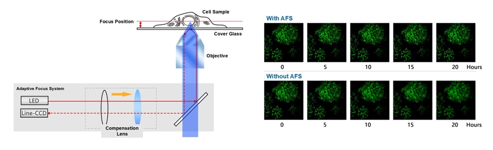
Even in a carefully controlled environment, focus drift is an invisible threat to long-term live-cell experiments. As heat from a stage-top incubator — or simply the ambient temperature of the room — transfers into the microscope body over time, minute thermal expansions shift the focal plane away from your sample. The result: a time-lapse sequence where the blur you see isn’t your cells moving, it’s your microscope drifting. Hours of imaging, compromised.
The EXI-1000’s Adaptive Focus System (AFS) eliminates that risk. Using a dedicated, independent focusing mechanism that operates continuously in the background, the AFS monitors and corrects the focal position in real time — compensating for thermal and vibrational disturbances before they ever appear in your images. Because the focus mechanism is isolated from other mechanical components, adjustments elsewhere on the microscope don’t disturb the Z-axis, giving you a stable foundation from the first frame to the last.
The practical benefit is simple but profound: every change you observe in a time-lapse sequence reflects what is actually happening in your sample — cells dividing, migrating, or responding to a treatment — not an artifact of instrument drift. Whether you’re imaging with a high-magnification, high-numerical aperture objective or pushing into advanced modalities like confocal, TIRF, or super-resolution, the AFS ensures your data is trustworthy, your experiments are repeatable, and your time is never wasted on a sequence you can’t use.