
Upright Microscopes

ACCU-SCOPE upright microscopes deliver top quality optics for bright, crisp images. Sometimes referred to as compound microscopes, our upright microscopes are favorites across a wide range of laboratories including clinical, veterinary, pharmaceutical, research, university, and education. Some of the disciplines utilizing our upright microscopes include pathology, hematology, microbiology, cytology, and histology. ACCU-SCOPE upright microscopes offer a selection of magnifications from ultra-low (e.g. 2X) to high power oil immersion (e.g. 100X). Our microscopes have a variety of options and accessories to perfectly fit your application, including ergonomic viewing heads, trinocular heads for camera attachment, and an assortment of objectives. Common contrast methods used with our microscopes include brightfield, phase contrast, darkfield, Differential Interference Contrast (DIC), and fluorescence.

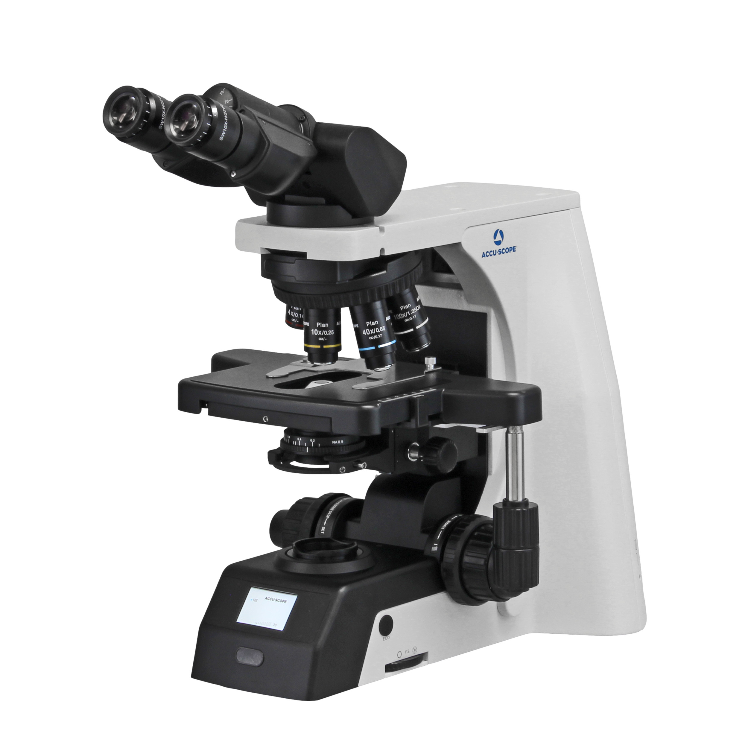
RC500

The ACCU-SCOPE RC500 remote collaboration system enables seamless automation and telepathology utilization, whether the user is sitting at the...
Learn More

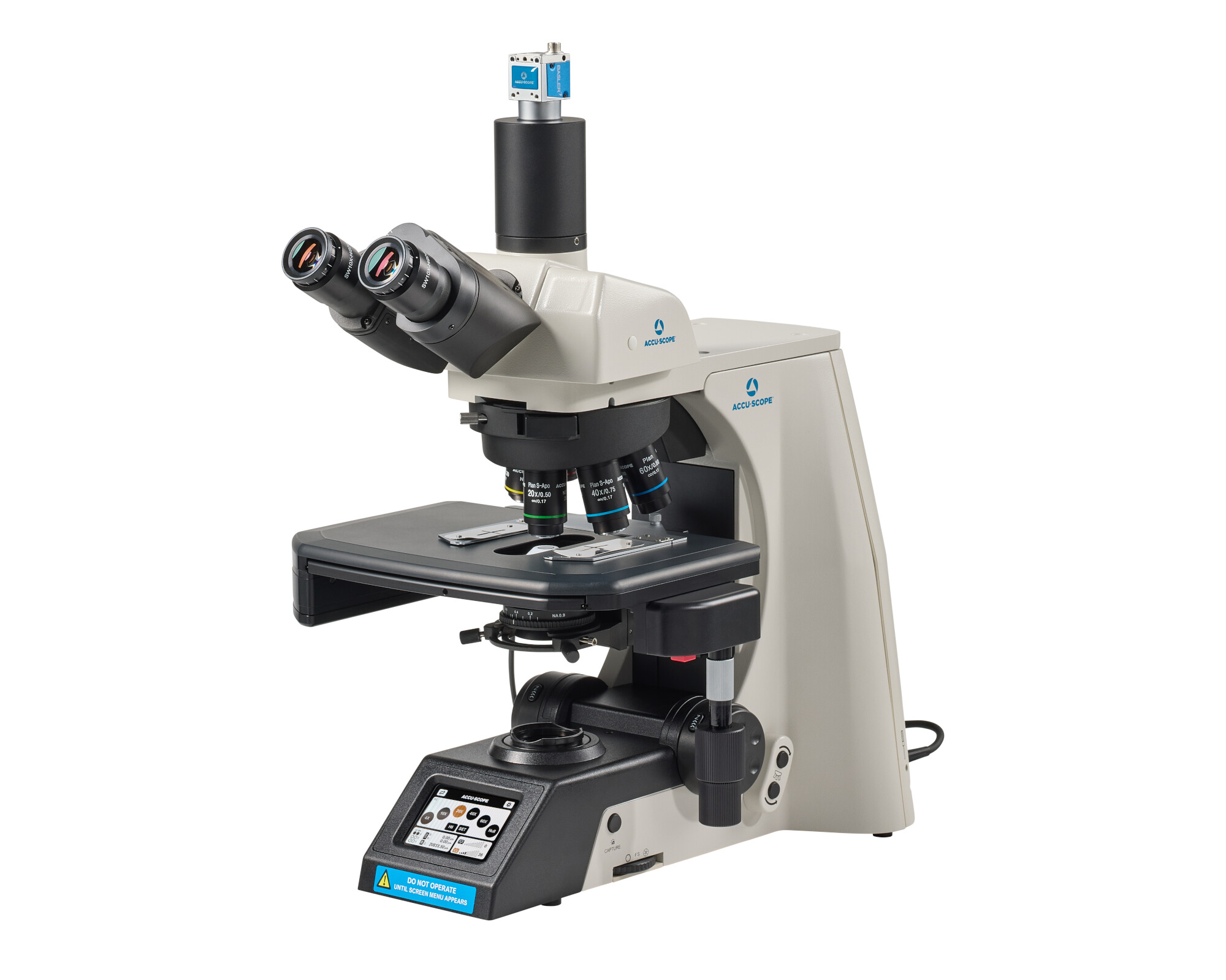
EXC-500

ACCU-SCOPE’s flagship EXC-500 Microscope offers best-in-class performance and value for clinical laboratory and research applications. The NIS...
Learn More

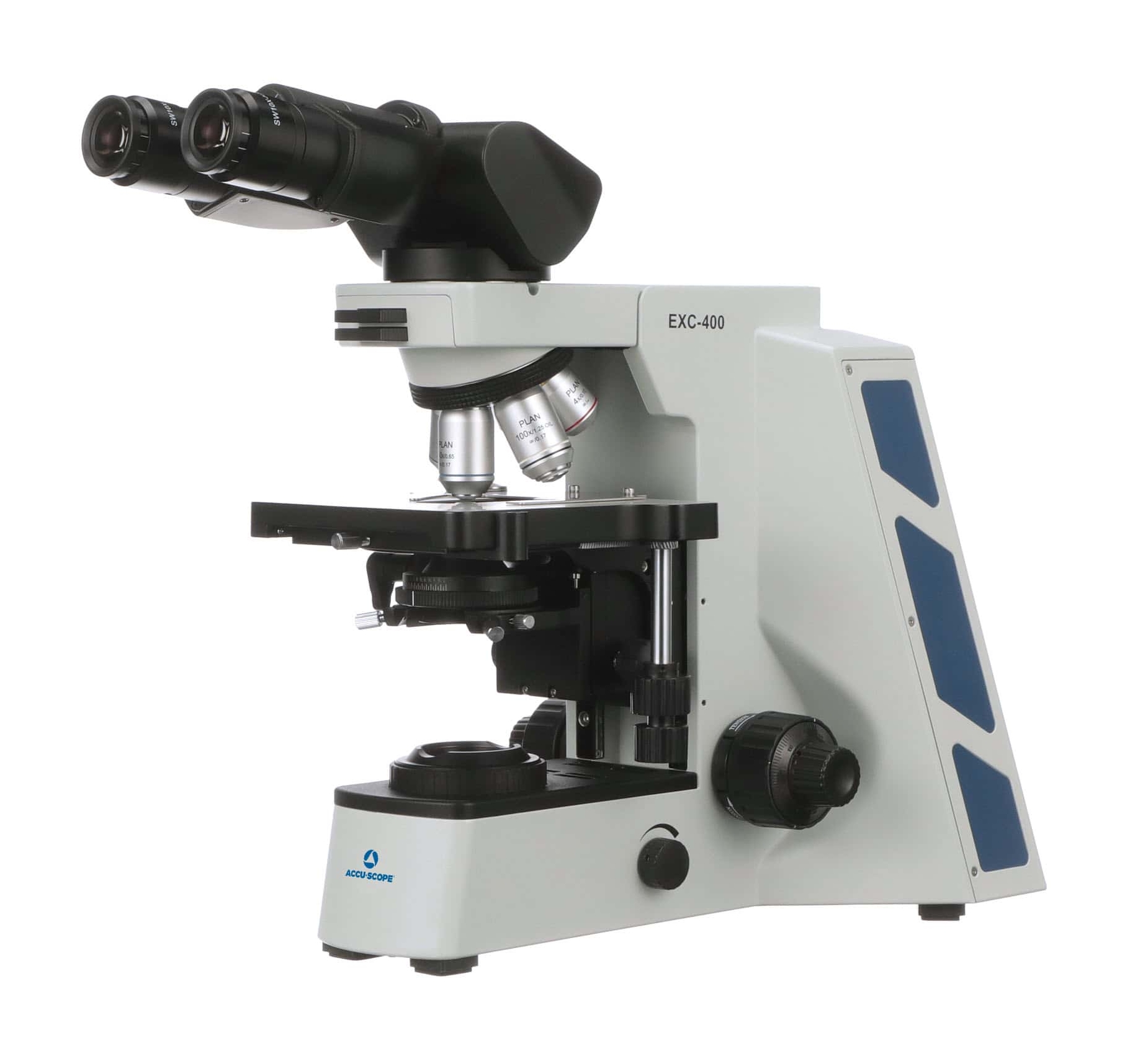
EXC-400

The EXC-400 from ACCU-SCOPE is designed for a broad range of microscopy applications in clinical, academic, and research environments. The EXC-400 is...
Learn More

What to Consider When Choosing an Upright Microscope

Choosing an upright microscope starts with the specimen and the work the laboratory performs most often. Magnification is important, but optical quality, stage movement, focus control, ergonomics, illumination, contrast methods, and digital imaging can all affect daily use. Comparing these features as a complete system can help narrow the upright microscope options that fit the application. Comparing microscopes by application keeps selection practical.

Optical Quality and Objective Selection

Objective selection should match the specimen and the detail the user needs to observe. Lower-power objectives provide a broader viewing area for locating material and reviewing larger structures. Higher-power objectives reveal finer detail but reduce the visible field. For many compound microscopes, a useful configuration includes several objectives so the user can move through different viewing magnifications during an examination. Objective choices vary across microscopes.

The optical system should be considered in its entirety rather than judged only by its highest magnification. Resolution, contrast, field of view, and image quality all affect the information the viewer can see. When comparing upright microscopes, look at the available objective choices and how they support the laboratory’s routine specimens and research needs.

Read more

Stage and Focus Controls

A mechanical stage allows the user to position a slide and move across the specimen in a controlled pattern. Smooth X-Y movement can be especially useful when a laboratory needs to scan a larger area of a slide. Stage controls are generally similar but may vary slightly between microscope models.  Coarse focus brings the specimen into view quickly, while fine focus supports smaller adjustments when working at higher magnification. 

Stage stability and precise focus become more important as magnification increases. The controls should be easy to reach and operate during routine work. When evaluating an upright microscope, consider stage travel, focus response, and how often users will need to reposition or scan specimens during a typical session.

Read more

Ergonomics and Viewing Configuration

Upright microscopes may be available with binocular, trinocular, or ergonomic viewing configurations. Binocular heads support direct observation through two eyepieces, while trinocular heads add an imaging path for a compatible camera. Ergonomic viewing heads may provide additional positioning options for users who spend longer periods at the microscope. These microscopes may support different viewing setups. 

Interpupillary adjustment, diopter settings, control placement, and viewing height can also affect how comfortably the microscope fits the user. Laboratories with multiple operators should consider how easily the system can be adjusted between users. These practical details can matter as much as optical specifications during repeated daily use.

Read more

Applications for Upright Microscopes 

Upright microscopes are widely used when specimens are viewed on slides placed on a stage beneath the objective lenses. In clinical laboratories, these microscopes support work in pathology, hematology, microbiology, cytology, and histology. Veterinary laboratories also use upright compound microscopes for slide-based examination and other prepared specimens. In research and university settings, an upright microscope can be a practical choice for tissue sections and biological samples that fit a traditional stage configuration. Model selection should reflect the specific needs of the laboratory. 

Education laboratories rely on upright microscopes for teaching students how to observe prepared slides and understand basic microscopy. The right configuration depends on the specimen, the level of detail required, and the type of work performed. A microscope selected for routine teaching may, however, call for different objectives, controls, and viewing options than one used for advanced clinical or research applications.

Illumination and Contrast Methods 

Illumination and contrast should match the specimen and the structures that need to be seen. Brightfield is a common method for many prepared and stained specimens. Phase contrast can improve visibility in certain transparent, unstained samples, while darkfield can make selected structures stand out against a dark background. Differential Interference Contrast (DIC) and fluorescence support more specialized applications when the microscope is configured for those techniques. Some microscopes support multiple contrast techniques. 

Not every upright microscope requires the same illumination or contrast options. The laboratory should identify its routine samples and methods before selecting a configuration. LED illumination, where available on the selected model, can provide a practical light source for routine observation and digital image capture.

Digital Imaging With Upright Microscopes

Digital imaging can extend an upright microscope beyond direct visual observation. A compatible camera can support specimen documentation, teaching, collaboration, and image review. Trinocular microscopes are often selected when a dedicated imaging path is needed while the eyepieces remain available for routine viewing. Camera-ready microscopes can support shared viewing. 

The microscope configuration should consider camera/imaging planning, adapter requirements, output or interface, and software used in the workflow. ACCU-SCOPE offers digital cameras and imaging solutions for clinical, veterinary, research, and educational microscopy. Considering future imaging needs during microscope selection can make it easier to add a camera later without replacing the core system.

Frequently Asked Questions About Upright Microscopes

What Is an Upright Microscope?

An upright microscope places the objective lenses above the specimen, with the sample positioned on a stage below. This traditional design is widely used for prepared slides and many biological samples. Upright microscopes are also commonly referred to as compound microscopes in laboratory and educational settings.

Upright microscopes support a broad range of clinical, veterinary, research, and educational work. Common uses include examining prepared slides, tissue sections, blood samples, microorganisms, and other specimens suited to an upright stage. The required objectives, contrast method, and viewing configuration depend on the specimen and the level of detail needed. These microscopes are common in slide-based work.

Phase contrast is useful for certain transparent or unstained specimens because it can make structures easier to distinguish without routine staining. It’s not required for every application. Laboratories should select phase contrast when it supports the specimens and observation methods used in their workflow or when required by examination protocols.

A camera can be added to compatible upright microscope configurations. Trinocular models provide an imaging path that can support a camera while preserving visual observation through the eyepieces. Camera compatibility depends on the microscope, adapter, interface, and software requirements, so future imaging plans should be considered when selecting the system. Many microscopes can support imaging with compatible hardware.

Choosing an ACCU-SCOPE Upright Microscope

When comparing upright microscope manufacturers, focus on the configuration that fits the specimen, application, magnification, contrast method, ergonomics, and imaging needs of your laboratory. Review the ACCU-SCOPE upright microscopes above or contact our team for application support and help to identify a system that combines optical performance, practical features, and strong value.