Posted on Leave a comment

ACCU-SCOPE to Exhibit at ACVP/ASVCP 2019

ACCU-SCOPE is excited to exhibit at the 2019 Concurrent Annual Meeting of ACVP and ASVCP in San Antonio, TX. On display and available for demonstration will be:

  1. EXC-500 upright microscope featuring semi-Apochromatic objectives, an ergonomic trinocular head and our ACCU-SlideMS manual slide scanning software.
  2. EXC-400 upright clinical microscope with side-by-side dual viewing accessory.
  3. EXC-350 upright microscope with single-channel fluorescence attachment (470nm for FITC/Alexa Fluor 488/GFP), a trinocular head and new Excelis MPX-6C 6MP color camera.
  4. 3013 trinocular upright microscope featuring an Excelis HDS camera with HD monitor.

Stop by Booth #201 and take any of our microscopes for a test drive. Bring your own slides, too!

Posted on Leave a comment

ACCU-SCOPE becomes Master Distributor for X-Cite

[October 2019] Effective October 1, 2019, ACCU-SCOPE Inc. becomes a Master Distributor in the US for X-Cite products from Excelitas Technologies Corporation.

“With the industry trend towards higher efficiency, cost effectiveness and environmental safety, plus our growing portfolio of fluorescence-ready microscopes, ACCU-SCOPE will focus our support on the new Mini+ and Xylis families of LED-based products,” said Brian Taub, Executive VP of ACCU-SCOPE. “By far, the Mini+ meets the needs of the vast majority of our customers. Where their applications require even higher intensity or longer wavelengths, the Xylis would be a better fit.”

The compact Mini+ delivers white light for fluorescence microscopy applications. With direct couplers for most microscopes, the only decision for a customer to make is whether they need a 380-700nm wavelength range or the extended 360-700nm version. The Mini+ supports use of common fluorophores such as DAPI, CFP, GFP/FITC, YFP, TRITC/Cy3/RFP/mCherry, MitoTracker® Red CMXROS and Cy5.

The Xylis is equipped with a liquid light guide and adapter to deliver white light to most fluorescence-ready microscopes. The Xylis rivals arc-lamp systems for brightness, and boasts wavelength range options of 380-770nm or 360-770nm, wide enough to support Cy7 in addition to all of the fluorophores for the Mini+.

ACCU-SCOPE dealers will be able to reserve demonstration systems for both the Mini+ and Xylis, so that customers can test them with their own samples and in their own laboratories. Contact your local ACCU-SCOPE dealer for more information about X-Cite illuminators for fluorescence microscopy, or contact ACCU-SCOPE at info.www.accu-scope.com to be referred to a dealer.


Posted on Leave a comment

Cleaning Microscope Optics

We all agree that a clean windshield or clean glasses work better and are more pleasing to look through. Microscopes work better when the optics are clean, too.  Dust and smears reduce the resolution, generate artifacts, and generally degrade the image, sometimes to the point of making the microscope useless.  Below are a few basic guidelines we use to clean microscope optics.  For best results, follow these steps in increasing order and NEVER soak the optic in any solvent!

  1. Use a puffer (Figure 1)
    • Remove surface dust by blowing air across it, and you only need a puffer or compressed air to do this.  This also avoids potential for scratching or smearing optical surfaces.
  1. Use lens tissue
    • Lens tissue has no lint, and lint can cause scratches.  We avoid using Kimwipes, too, as they may be abrasive.  Gently wipe an optical surface in a circular pattern, beginning in the center and spiraling outward (Figure 2).  Repeat. FYI, facial tissue contains a wetting agent that may add smears to the glass surface.
  1. Use water or warm breath
    • In combination with Step 2, a warm breath may be sufficient to remove the dust, debris, and some smears.  You may also wet the lens tissue slightly before wiping.  We will also wrap the lens tissue around a cotton-tipped applicator stick or Q-tip.  Alternatively, you can fold the lens tissue into a point (Figure 4a; courtesy of https://micro.magnet.fsu.edu).
  1. Use alcohol or lens cleaner (WEAR NITRILE GLOVES)
    • Alcohol or lens cleaning solution (Figure 3) is the next solvent after water/breath.  One of our favorites is Sparkle glass cleaner, but dilute this 1:1 with distilled or deionized water (a.k.a. Milli-Q water).  70% isopropanol or 70% ethanol is also acceptable, but not denatured alcohol.  Be careful when cleaning the eyepieces as these solvents may damage the rubber eyeshields.  Wrap lens tissue over applicator stick or Q-tip as above, and slightly dampen with solvent before wiping.  Work in circular/spiral motion from the center outward.  Repeat.
  1. Use stronger solvents (WEAR APPROPRIATE GLOVES), or seek professional help
    • In some cases, and with some stuck-on substances like dried-on immersion oil, you may need to use other solvents such as xylol or a 1:1 mixture of ether-ethanol.  IMPOTRANT! DO NOT SOAK THE OBJECTIVE IN THE SOLVENT as this may soften the cement securing the lens elements.  Other organic solvents may also be used for more stubborn crud (Residual Oil Remover, pure petroleum ether, etc.).  Follow same procedure as in Step 4, and use sparingly!

One final tip.  It is easier to inspect and clean objectives if they are removed from the microscope.  Remove one of the eyepieces and look through it the “wrong way” (e.g. from the back; Figure 4b) at the lens – – it’s a handy 10x magnifier!  With a few seconds of practice, you can see smears, dust and even damage to the lens.

Enjoy your clean microscope and, to maintain peak performance, remember to clean those optics on a regular basis.

Posted on Leave a comment

Demystifying Numerical Aperture

We are often asked to recommend the objectives for a microscope.  The objective sits closest to the specimen, and is an integral component of the microscope and crucial to delivering an acceptable image of the specimen.  All the information you need to know is written right on the barrel of the objective: flatness correction, magnification, Numerical Aperture, immersion medium, optical path design, and whether to use with or without a coverglass.  One of the more confusing criteria, however, is Numerical Aperture.  In this brief article, I’ll attempt to demystify Numerical Aperture, and how to get the most out of it.

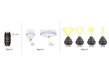
Very simplistically, Numerical Aperture (“NA”) is the ability of an objective lens to collect light, and it’s written on the objective just after the magnification (Figure 1).  A higher angle of light = a higher NA (see Figure 2).  Additional light means more information, which means better resolution.  So the higher the NA, the better the optical resolution.  But it’s not as easy as moving closer to the specimen.

In order to take maximum advantage of the objective’s NA, the condenser should be adjusted to match – this adjustment is one step of Köhler Alignment.  Thankfully most condensers have an aperture adjustment for this purpose (see Figure. 3).  Since the NA changes every time you change objectives on a microscope (objectives with higher magnification typically have higher NA), the condenser should also be adjusted – this is literally a 2 second adjustment, and can make a HUGE difference in the image and resolution.  We’ll review Köhler Alignment in another article.

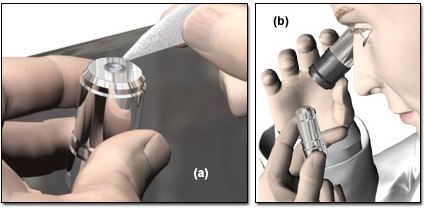
Let’s say that two objectives have the same magnification but different NAs. The one with higher NA will typically cost more.  And to get an NA of 1.0 or higher, you’ll need an immersion objective that requires some other medium than air (typically oil, water, glycerin, or silicon oil) to be placed between the objective and the coverglass above the specimen.  This additional medium bends (“refracts”) more light (therefore, more information) into the objective lens, thereby increasing resolution. Note that immersion objectives are specifically designed for particular immersion media, and no objective should be used with an immersion medium for which it was not intended – – this will void any warranty, and you won’t get the results you hoped for.

Numerical Aperture = n sin θ

where n is the refractive index of the medium between the objective and the coverglass, and θ  is the ½ angle of light collected by the objective lens (refer to Fig. 2).  Air has a refractive index of approximately 1.0 and typical immersion oil has a refractive index of 1.51.  You can see how oil is needed for an NA > 1.0.  Therefore in order to have an NA greater than 1.0, you’ll need to use an immersion medium with a refractive index higher than 1.0.

Objectives of the same magnification but different NAs will give different results.  A higher NA objective, when the microscope is properly adjusted, will have higher resolution and deliver a crisper image.  The trade off is that the depth of focus becomes shallower as resolution increases.  So if you want to have more of the specimen in focus, you may want to choose an objective with lower NA.

Finally, higher NA doesn’t necessarily translate into a better image.  Other optical qualities must be considered (e.g. correction for field curvature, chromatic aberration, spherical aberration, etc.), all topics for another time.

Thanks for reading!

Posted on Leave a comment

ACCU-SCOPE Introduces CaptaVision+ for Excelis Cameras

The new CaptaVision+ software features a new customizable user interface with several new features including real-time image stitching, real-time depth-of-field fusion, report generation and export, and more!

CaptaVision+ ships with the following Excelis camera models:

  • Excelis HD and HDS
  • Excelis MPX-5C Pro
  • Excelis MPX-6C

Excelis HD Lite, HDS Lite and MPX-6C will continue to use CaptaVision v5.1.

Stay tuned for a series of video tutorials to help you get the most out of the new CaptaVision+!

Posted on Leave a comment

NEW Excelis™ MPX-5C Pro and MPX-6C Microscopy Cameras

[April 2019] The Excelis™ 5MP MPX-5C Pro and 6MP MPX-6C deliver exceptional performance in a compact, low-profile design and are the latest additions to our Excelis line of microscopy cameras! The revolutionary, feature-rich CaptaVision+ software provides real-time image stitching, real-time depth-of-field fusion, report generation & export, and more!

The MPX-5C Pro features a 5MP color camera with SONY IMX264LQR-C CMOS sensor, ultra-high speed USB 3.0 connectivity, 2448 (H) x 2048 (V) resolution at 35FPS, global shutter; exposure time 0.13ms-15s, and CaptaVision+ imaging software with real-time depth-of-field fusion, real-time image stitching, real-time fluorescence image synthesis and editing & more!

The MPX-6C Camera features a 6 MP color camera, SONY IMX178LQJ-C CMOS sensor. ultra-high speed USB 3.0 connectivity, 3072 (H) x 2048 (V) resolution at 40FPS, rolling shutter; exposure time 0.13ms-15s, an CaptaVision+ imaging software with real-time depth-of-field fusion, real-time image stitching, real-time fluorescence image synthesis and editing & more!

“Click to learn more about the Excelis MPX-5C PRO and MPX-6C.”

Posted on Leave a comment

The NEW EXC-400 Microscope Hits the Mark for Clinical and Research Labs

[June 11, 2018] — ACCU-SCOPE Inc. is pleased to introduce the EXC-400 Microscope, the latest addition to its growing portfolio of microscopes specifically designed for clinical and research applications.  The EXC-400 redefines quality, durability, convenience and efficiency for a broad range of microscopy applications in clinical, academic and research environments.

The EXC-400 is ideal for routine observation of specimens but is, itself, far from routine.  Standard features of the EXC-400 include a trinocular viewing head, a wide 22mm view through the eyepieces, a ceramic hard-coated stage for maximum durability with repeated use, a quintuple nosepiece for holding up to five objectives, infinity corrected optics to meet the latest demands of technicians and researchers, and LED illumination for dependable cost-effective operation for years of worry-free use.  The EXC-400 can be configured for several contrast methods including brightfield, phase contrast, polarized light, Gout analysis, and fluorescence featuring integrated LED illumination and Chroma® filters.  The standard Plan Achromat objectives provide excellent color reproduction and resolution of details, and the optional semi-Apochromatic objectives are an excellent choice especially for fluorescence observation and imaging.  An optional dual observation attachment allows two people to view the specimen simultaneously, perfect for discussion and training.

Capturing images with microscopes is nearly ubiquitous and, with its trinocular viewing head, the EXC-400 is ready to attach any one of the digital microscopy cameras from ACCU-SCOPE including the ExcelisTM and Lumenera® INFINITY series cameras.  “We are delighted to deliver the new EXC-400 to technicians, clinicians, and researchers in the hospital, clinical and research labs,” says Brian Taub, Executive VP at ACCU-SCOPE.  “With an impressive list of standard features, plus a long list of optional components and capabilities, the EXC-400 is ready to meet the demands of these discriminating scientists.”


Click HERE to learn more about the EXC-400.”


Posted on Leave a comment

NEW Videos from ACCU-SCOPE

[January 2019] After over six months of writing, filming and editing, we’re excited to share the final production of three videos that introduce ACCU-SCOPE, as well as two of our newest products, the EXC-400 and the EXC-500 microscopes.

Below you will find the video subject, a link to view the video, and embedding code.  Click the thumbnail image or the video link to view. You should also be able to download the video directly from Vimeo.  The videos are only a couple minutes long and provide an informative and compelling glimpse into our corporate identity, history and brand.

ACCU-SCOPE Company Video


EXC-500 Product Explainer Video


EXC-400 Product Explainer Video

View this an other product videos HERE.

Contact info@www.accu-scope.com with any questions.

Posted on Leave a comment

NEW WiFi-enabled Microscope Camera Adds Flexibility, Versatility and Convenience

[July 2018] — ACCU-SCOPE Inc. is pleased to introduce the SKYE WiFi™ microscopy camera.  The SKYE WiFi camera brings researchers and educators a new level of convenience and flexibility across a broad range of microscopy imaging applications for documentation, analysis, education and collaboration.

Utilizing the existing WiFi system in most facilities together with a mobile app for Android and iOS devices, the SKYE WiFi camera streams live images at up to 40 frames per second.  The user controls the camera with a WiFi-connected device, while the images can be simultaneously viewed on up to ten WiFi-connected devices.  No WiFi?  No problem.  The SKYE WiFi camera also features a USB mode for image acquisition and viewing through a PC using the included SKYE View™ software.  Upgrade almost any brand of microscope by attaching the SKYE WiFi camera with an industry-standard c-mount adapter, and the 5-megapixel camera sensor will deliver brilliant images with amazing resolution and detail.  The SKYE WiFi camera creates a new level of versatility for microscopy imaging for the laboratory and classroom.

Although the use of digital cameras with microscopes is quite common, not every laboratory has room for a computer by the microscope.  “We are delighted to deliver the new SKYE WiFi camera to technicians, teachers, and researchers across industries, applications and institutions,” says Brian Taub, Executive VP at ACCU-SCOPE.  “Couple the mobile device app with its built-in WiFi connectivity, and the SKYE WiFi camera is the perfect complement for instruction, documentation and collaboration by today’s discriminating scientists and educators.”

Click HERE to learn more about the SKYE WiFi camera.