Veterinary

Microscopes are indispensable instruments in veterinary clinics, allowing for quick examination of samples for abnormalities and providing valuable diagnostic observations. Common uses of microscopes in veterinary laboratories include Urinalysis, Parasitology, Microbiology, Cytology, Checking fecal samples for parasites, and Diagnosing skin conditions. Microscopes are used daily to view a variety of biological substances, including: blood, urine, fecal material, fluid aspirates, skin scrapings, and ear cerumen.

ACCU-SCOPE offers a range of microscopes that are ideal for the veterinary laboratory. Many of our microscopes readily accommodate the addition of a digital camera, ideal for documentation and training.

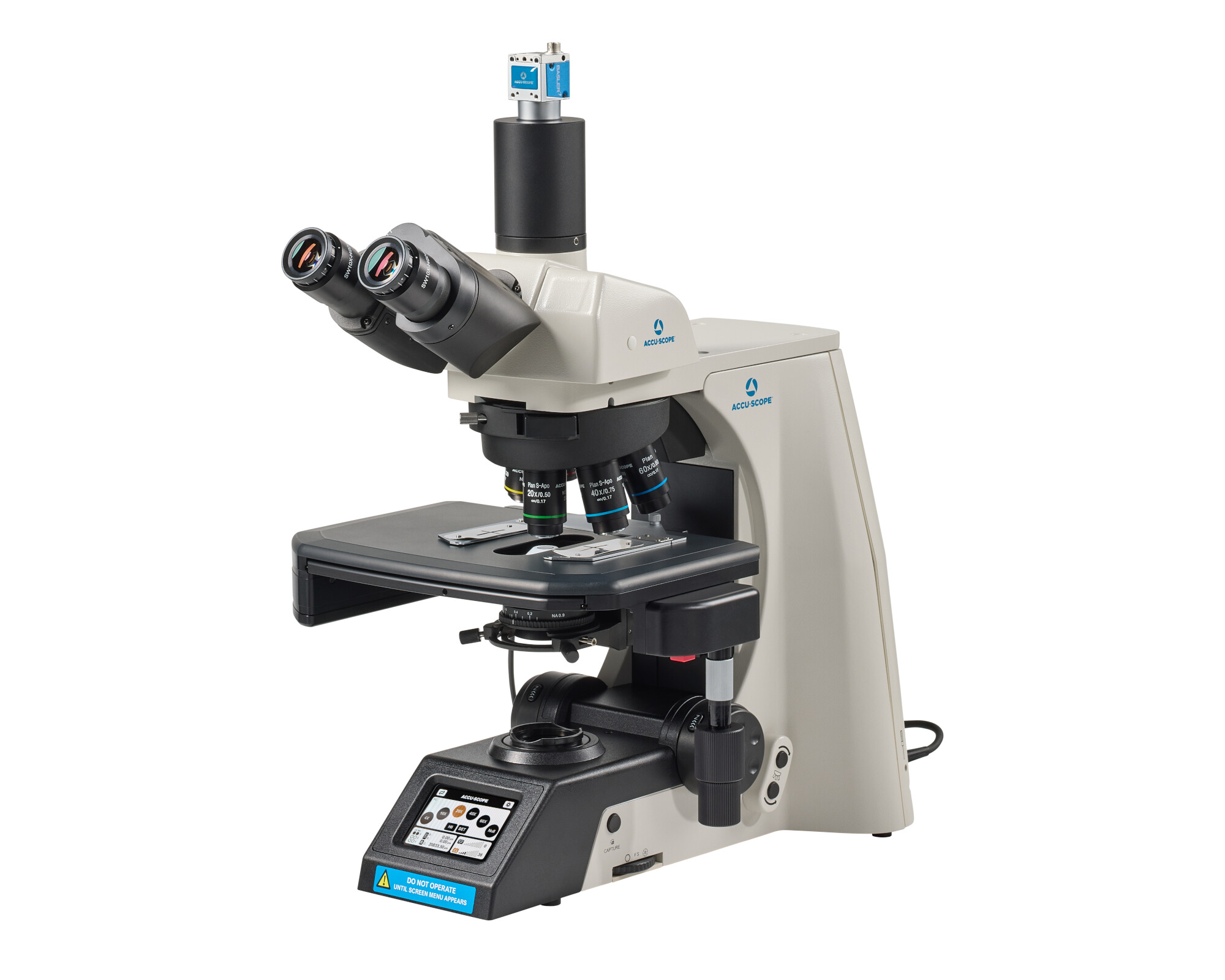
RC500

The ACCU-SCOPE RC500 remote collaboration system enables seamless automation and telepathology utilization, whether the user is sitting at the...
Learn More

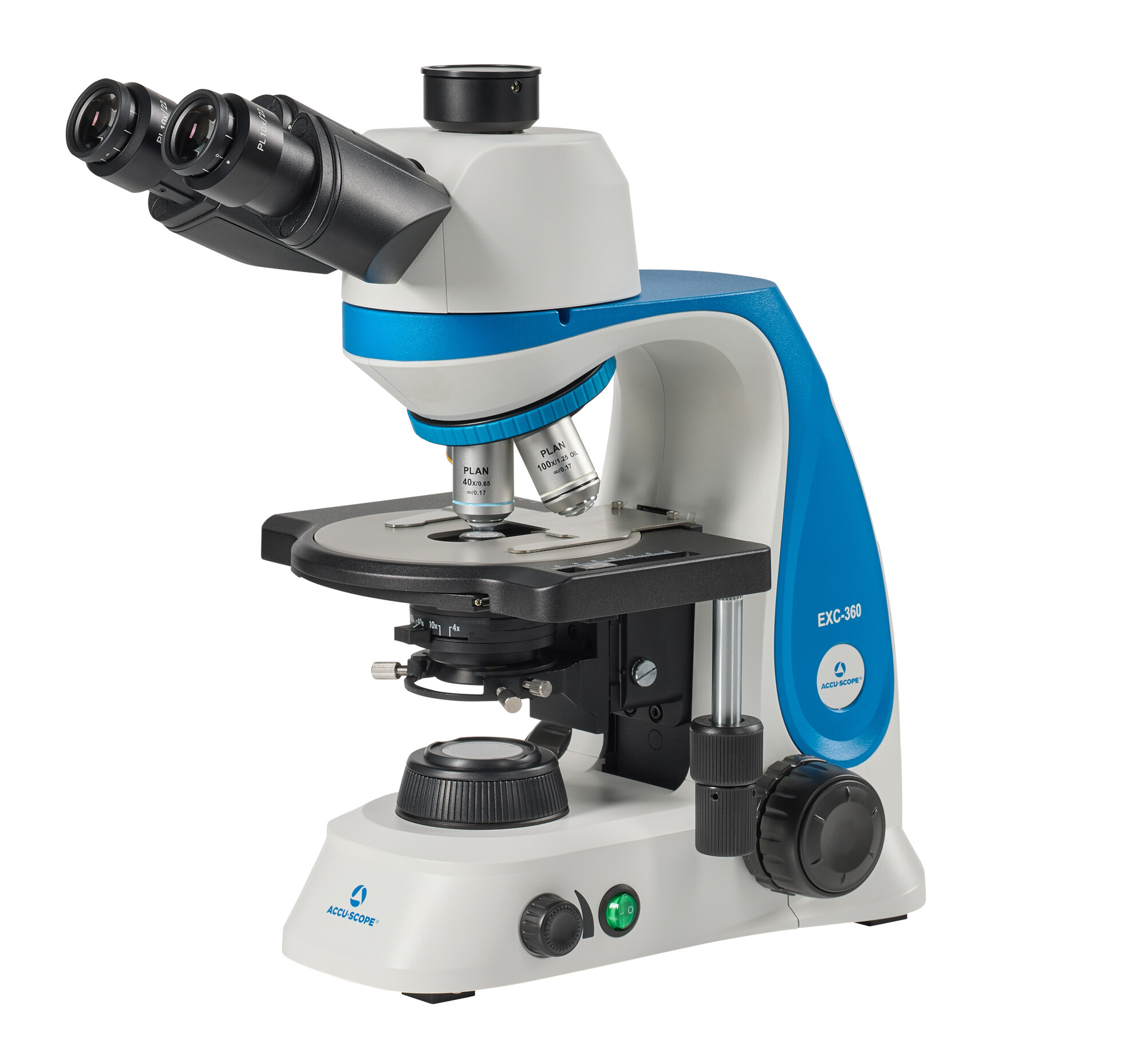
EXC-360

The NEW EXC-360 from ACCU-SCOPE delivers exceptional optical performance, versatility and durability for clinical, veterinary and education...
Learn More

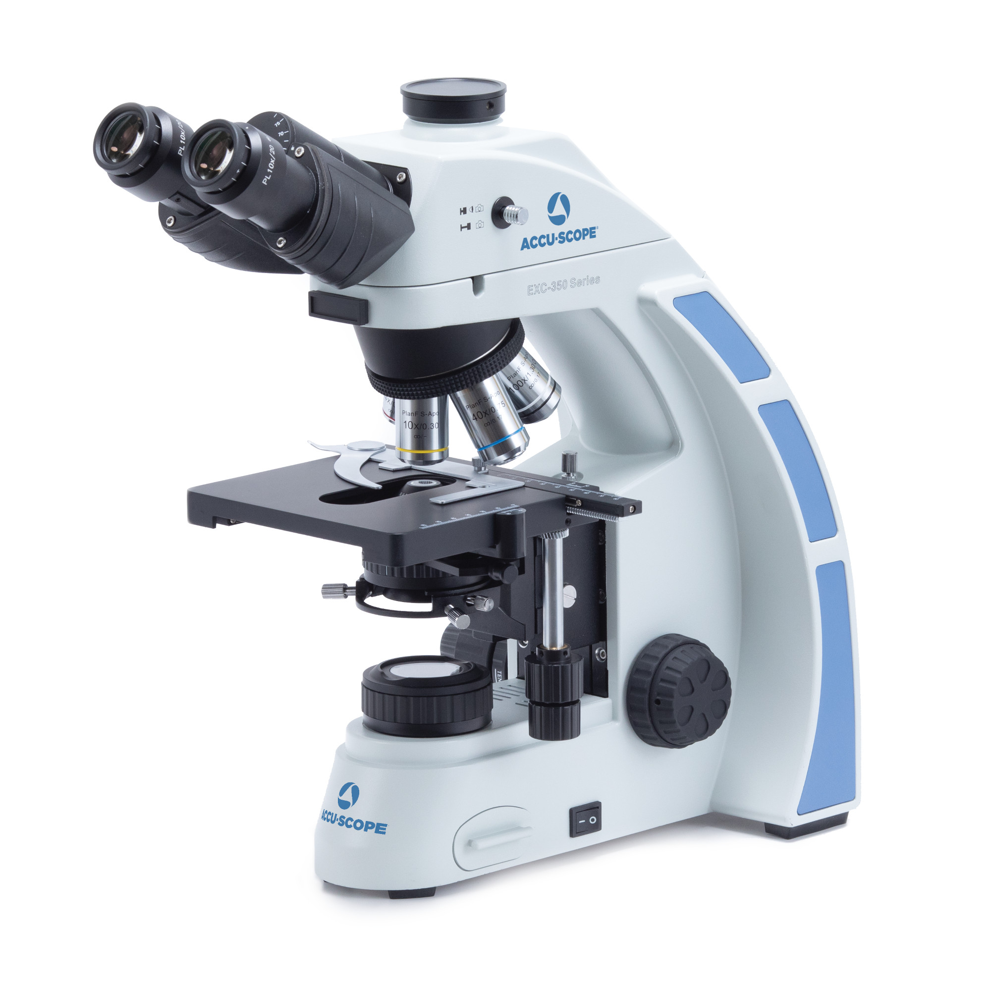
EXC-350

The EXC-350 from ACCU-SCOPE delivers a high level of optical performance, versatility and durability for clinical, veterinary and education...
Learn More

Veterinary Microscope Options for Clinics and Labs

When comparing veterinary microscopes, clinics need to consider their specimens, required objective lenses, and number of users. High-quality optics, simple controls, digital imaging, and straightforward maintenance support microscopes used across veterinary clinics, laboratories, classrooms, and field settings.

How Microscopy Supports Veterinary Workflows

Microscopy provides a direct view of cells, organisms, and other structures. Microscopes are essential for investigating animal health conditions through fecal exams, urinalysis, cytology, and blood-smear review. The right microscope can identify parasites in fecal samples. Urinalysis examines sediment for evidence related to urinary and systemic conditions, and cytology uses fine-needle aspirates to investigate disease. Findings should always be taken into consideration along with the medical history, physical examination, and other tests.

Busy teams save time by preparing supplies, labeling specimens, and following the same viewing sequence. Starting at a lower power helps users locate the right area before changing objective lenses. Consistent illumination, focus, and slide handling help make images easier to compare. Fresh urine samples, properly prepared smears, and clean skin scrapings give staff stronger starting points, too.

Microscopic findings can support or confirm a diagnosis and track treatment progress. Additionally, they support veterinary science education and research. Cytology can guide further testing, while microscopic review before a procedure can inform surgical planning and contribute to better outcomes. Specialty laboratories sometimes use fluorescence microscopes to detect selected pathogens or markers that inform treatment planning.

With trained staff and suitable use cases, in-house microscopy can reduce outside testing costs and turnaround time

Choosing a Microscope for Veterinary Applications

Compound microscopes are standard for many tasks in veterinary medicine since they provide the magnification needed for prepared slides. A microscope veterinary professionals use for routine work commonly includes 4x, 10x, 40x, and 100x objective lenses. High-quality optics are crucial for distinguishing fine detail, and mechanical stages control slide movement.

Stereo microscopes offer lower magnification, a three-dimensional view, and more room for specimens. Veterinary microscopes can have monocular, binocular, or trinocular heads. Binocular microscopes support comfortable viewing, while trinocular veterinary microscopes add a camera path.

Brightfield observation is also common for routine slide work. LED microscopes use less energy, with their lamps lasting longer than many traditional sources. Halogen microscopes, however, remain preferred where bright, concentrated light is needed.

For more frequent use, select microscopes with accessible focus controls, easy-to-clean stages, durable construction, and ergonomic viewing. High-quality optics are invaluable when staff share the same microscopes throughout the day.

Field teams should prioritize size, stable illumination, power, carrying protection, and setup time. Cordless or rechargeable LED microscopes can be advantageous where outlets are limited, but they still require secure transport and a stable surface.

Digital Imaging, Documentation, and Remote Collaboration

Adding a camera allows veterinary clinics to capture images for documentation, training, comparison, and consultation. Trinocular microscopes can accept a camera while keeping the eyepieces available.

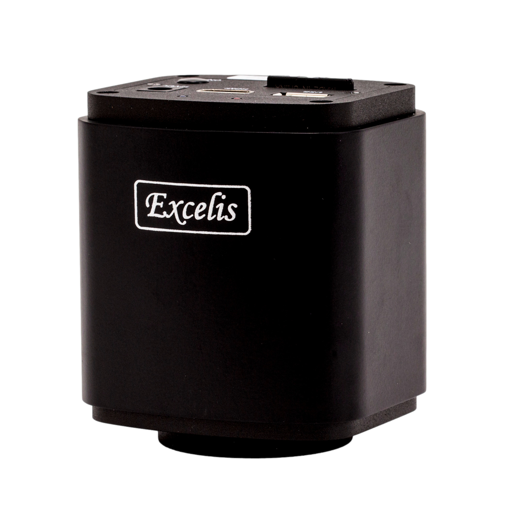
Resolution is an important selection factor, as well as sensor size, color, frame rate, software compatibility, and display. The Excelis HD camera provides 1080p live imaging, captures 6 MP images, and comes with onboard measurement and annotation tools.

Imaging software helps capture, annotate, measure, and manage images from microscopes. These tools can mark areas of interest and build archives for teaching or reference; clinics should name files with the case ID, sample type, and collection date, then store images under their record, access, and retention policies. CaptaVision+ supports these functions with compatible Excelis cameras.

The RC500 Remote Collaboration System lets remote users control the stage, focus, objectives, and illumination, and they can also capture, annotate, and measure images. Remote operation requires a strong internet connection, and captured images still need thorough interpretation.

EXCELISâ„¢ HD

The Excelisâ„¢ HD camera delivers true, live 1080P HD images at 25fps, captures 6MP photos and records live video in full HD. On-board software provides measurement, annotation, comparison and control over exposure, gain and other camera settings without a computer. The AU-600-HDS version includes an attachable HD monitor that can be mounted directly to the…

View product

CaptaVision+ Software

CaptaVision+â„¢ is our powerful software package, exclusively developed to integrate with our Excelisâ„¢ line of microscope cameras. Featuring a logical workflow, CaptaVision+ provides for precise camera control, image acquisition, image management, image processing, measuring and counting. CaptaVision+ v3.1 adds new versatility and power for image acquisition, processing and analysis.  CaptaVision+ v3.1 is available for Windows…

View product

RC500

The ACCU-SCOPE RC500 remote collaboration system enables seamless automation and telepathology utilization, whether the user is sitting at the microscope or located remotely. The RC500 system allows the remote user to select the objective magnification, move the stage to reposition the specimen, adjust illumination intensity, capture, annotate and measure images. The RC500 comes complete with…

View product

Microscope Care, Accessories, and Staff Training

Routine maintenance protects microscopes, keeping them ready for everyday use. Cover microscopes when idle, clean lenses with approved materials, and promptly remove immersion oil. Check the microscope cords, stage controls, and illumination consistently, and arrange professional service according to use and manufacturer guidance.

Essential supplies include immersion oil, lens paper, approved cleaning solution, slides, coverslips, and the correct camera adapter. Additionally, keep spare lamps for microscopes with replaceable bulbs. Train all new users on setup, focusing, oil immersion, shutdown, cleaning, and image procedures, with refreshers on written veterinary microscopy protocols.

Frequently Asked Questions About Veterinary Microscopes

What objective magnifications are commonly used in veterinary microscopy?

Many compound veterinary microscopes use 4x, 10x, 40x, and 100x objectives. Lower powers help locate and scan a sample, while higher powers show finer detail. With a 10x eyepiece, a 40x objective gives 400x total magnification; a 100x objective gives 1,000x.

A 100x oil objective is used when the protocols call for greater resolution of small cellular or microbial detail. Apply the oil only to objectives designed for immersion. After viewing, remove it promptly with approved lens-cleaning materials.

Compound microscopes provide higher magnification for thin specimens. Stereo microscopes offer lower magnification, a three-dimensional view, and more room below the objective. The right choice is shaped by the sample and the work performed by the veterinary practice.

Digital cameras can capture images from microscopes for records, measurements, training, comparison, and consultation. They also let several people view one field on a monitor. Digital images support the record, but they don’t eliminate the need for qualified examination and interpretation.

Many cytology preparations are stained to improve contrast through microscopes. The stain and preparation method are dependent on the sample, examination, and laboratory protocol. Veterinary medicine teams should follow all validated procedures and request specialist review when a case falls outside their experience.

Ask about warranty length, covered parts and labor, exclusions, repairs, turnaround, and technical support for all microscopes and cameras. ACCU-SCOPE provides a five-year limited microscope warranty for qualifying products, with separate terms for LED lamps and other components. ACCU-SCOPE can help you compare veterinary microscopes, camera options, and imaging software. Contact our team today to discuss your needs for your clinic or laboratory.