
Dissecting Microscopes for Research and Laboratory Use

Dissecting microscopes give researchers, instructors, and laboratory professionals a clear, three-dimensional view of larger specimens at low-to-moderate magnification. Their broad field of view and generous working distance make them well suited for dissection, specimen handling, plant and animal anatomy, biomedical-device inspection, mineral examination, and quality-control work.

ACCU-SCOPE manufactures professional microscopy and imaging solutions for research, education, clinical, and industrial settings. As part of ACCU-SCOPE’s broader selection of research microscopes, these dissecting microscopes combine optical performance, practical configurations, and strong value. Explore the available models below to find the right combination of optics, working distance, illumination, and imaging capabilities for your application.

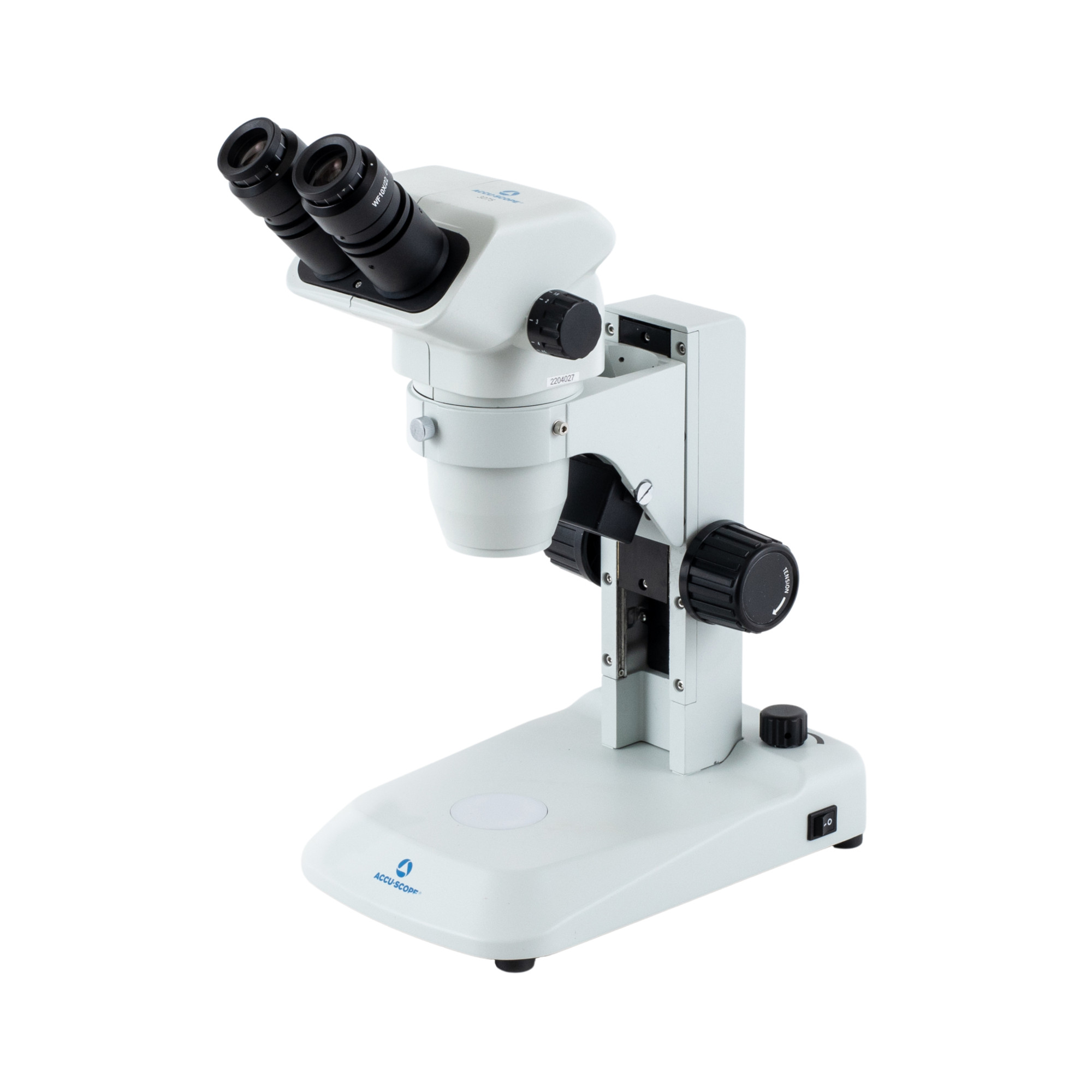
3075

The ACCU-SCOPE 3075 Series Zoom Stereo Microscopes are easy to use. Featuring a 0.67x – 4.5x zoom range, the 3075 Series delivers sharp, high...
Learn More

What Is a Dissecting Microscope?

A dissecting microscope, sometimes called a dissection microscope or stereo microscope, uses two separate optical paths to provide depth perception and a three-dimensional view of the specimen. This design helps users examine surface features while keeping enough space beneath the objective lenses for tools, forceps, containers, and hand movement. Unlike systems built mainly for thin slides, dissecting microscopes are often used with larger, opaque specimens that need little sample preparation.

Low-to-moderate magnification, a wide field of view, and a long working distance support detailed observation and manipulation. Applications include biological dissection, plant and animal anatomy, classroom study, biomedical-device inspection, mineral examination, and quality control, all of which benefit from a stable magnified image with visible depth.
Dissecting Microscopes vs. Compound Microscopes

Dissecting Microscopes

Dissecting microscopes and compound microscopes serve different viewing needs. A dissecting microscope is generally the better choice for larger specimens, surface examination, and tasks that require tools beneath the optics. It provides a three-dimensional view and typically relies on reflected light to illuminate the specimen from above. Transmitted light may also be useful when a sample allows light to pass through it.

Read more

Compound Microscopes

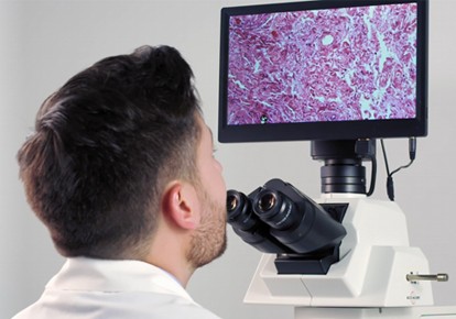
A compound microscope is designed for smaller, thin, or transparent specimens that require higher magnification. Its objective lenses sit much closer to the slide, which creates less room for manipulation. A compound microscope also produces a flatter view than a stereo system. For cell structures, prepared tissue sections, and many microorganisms, a compound microscope is usually the more appropriate instrument. For dissection and surface inspection, dissecting microscopes provide the working space and depth perception users need.

Read more

How to Select the Right Dissecting Microscope

The best configuration depends on the specimen, the task, and the need for image capture or shared viewing. Consider sample size, surface characteristics, tool movement, and required detail before comparing optics, stands, illumination, and imaging accessories.

Magnification, Optics, and Field of View

Zoom dissecting microscopes let users adjust magnification as specimen size or viewing needs change. They suit varied samples and tasks that move between broad inspection and finer detail. Fixed-magnification systems can support repeatable work at the same viewing level.

The optical configuration also affects field of view, image quality, and working distance. Objective lenses with lower power generally provide a wider viewing area and more room beneath the microscope. Higher power reveals finer detail but narrows the field of view. Beginning at a lower zoom setting makes it easier to locate the area of interest before increasing magnification. Eyepiece choice and objective lenses should be evaluated together so the full system matches the intended task.

Read more

Working Distance and Stand Configuration

Working distance is the space between the lowest point of the objective and the specimen when the image is in focus. Dissection tasks often need room for forceps, probes, specimen dishes, or other tools. Larger samples may also require added vertical clearance or greater reach across the work surface.

A stable benchtop stand is a practical choice for routine laboratory use with specimens that fit within a defined stage area. Pole stands support straightforward vertical adjustment. Boom stands extend the microscope head over larger samples and open work areas, making them useful when the specimen cannot sit on a standard stage.

Read more

Illumination for Opaque and Translucent Specimens

Reflected light directs illumination onto the specimen surface and is commonly used for opaque samples. Transmitted light passes upward through a sample and may be needed for translucent specimens or fine structures that benefit from backlighting. Some applications call for both types so the user can adjust the lighting as the specimen changes.

LED illumination is a practical option for long service life, limited heat, and stable viewing conditions. A cooler light source can also reduce heat exposure during longer observation sessions. The light should provide even coverage without creating glare that hides surface detail.

Read more

Ergonomics and Accessories

Microscope configuration affects posture and comfort during extended sessions. Adjustable eyepieces, tilting heads, focus-tension controls, and movable workstations can support a more natural viewing position. Stage plates and specimen holders may also improve control. Match accessories to the user, workstation, specimen, and session length.

Read more
Feature image

Digital Imaging and Documentation Options

Digital imaging can support research records, classroom instruction, collaboration, and comparison. HDMI cameras allow direct monitor viewing, while USB cameras connect to a computer for image capture and software-based work. Integrated displays may suit demonstrations or work areas without a separate computer.

When comparing camera options, consider resolution, frame rate, output type, capture software, and operating-system compatibility. The microscope should also have a suitable imaging path and adapter options. Even when a camera will be added later, planning for digital imaging during microscope selection can make future expansion easier.

Find the Right ACCU-SCOPE Dissecting Microscope

The right microscope should fit the specimen, viewing range, working distance, illumination, stand, and documentation needs. Review the ACCU-SCOPE products above, contact our team for application support, or connect with an authorized distributor to discuss a configuration that provides reliable optical performance and strong value.

Frequently Asked Questions About Dissecting Microscopes

What Is a Dissecting Microscope Used For?

A dissecting microscope is used to view and manipulate larger, three-dimensional specimens at low-to-moderate magnification. Common applications include biological dissection, anatomy study, specimen sorting, device inspection, mineral examination, and quality-control work.

A dissecting microscope, also called a stereo microscope, is commonly used for dissection. Its two optical paths provide depth perception, while the wide field of view and long working distance leave room for tools and specimen handling.

A dissecting microscope is designed for larger specimens, surface viewing, and hands-on manipulation. A compound microscope uses higher magnification for thin or transparent specimens, such as prepared slides. The compound microscope has a shorter working distance and does not provide the same three-dimensional view.

Reflected light is usually selected for opaque specimens because it illuminates the surface. Transmitted light is useful for translucent samples that allow light to pass through. Some dissecting microscopes support both illumination methods for greater application flexibility.

Measure the height of the specimen, container, and tools used during the task. Choose enough working distance to move comfortably without contacting the objective lenses. Larger samples and active manipulation generally require more clearance.

Many dissecting microscopes can be configured for digital imaging. Compatibility depends on the microscope head, camera mount, adapter, output, and software. Consider future camera needs when selecting the microscope, even when imaging will be added later.