Depth of field and depth of focus are both well-known terms in photography, filmography, and microscopy. Given how similar their names are, they have sometimes been used interchangeably. However, despite both playing a vital role in camera and microscope optics, the two are very much different. Let’s look more closely at what defines the depth of field and depth of focus, their role in microscopy, and why the two should not be confused with one another.
Depth of Field
The depth of field is the distance between the closest object plane in focus and the furthest one in focus. In other words, it can be considered a lens’s ability to retain the quality of an image without moving the object farther or closer to the frame. Depth of field relates to how much detail can be seen above and below the optimal focus position. When objects are placed at a set focal distance away from a lens, detail can blur and thereby degrade the resolution. As a result, smaller objects naturally lead to reduced depth of field for the lens. It is worth noting that the depth of field will increase as the condenser numerical aperture (condenser aperture diaphragm) is closed down (approaches zero). While depth of field increases when the condenser aperture closes, resolution decreases.
Depth of Focus
Depth of focus is the imaging complement to depth of field and is more concerned with the tilt of the sensor plane of the microscope with respect to the lens image plane. This concept relates to how the quality of focus is altered as the object stays still and the lens’s sensor side changes. Therefore, the depth of focus will change significantly based on the magnification of the object and the microscope’s ability to focus light, known as the numerical aperture. Depth of focus has little to do with sensor size and is more closely related to sensor perpendicularity to the optical axis and pixel count — the more pixels the sensors have, the more noticeable sensor tilt will be on the quality of the image. Given how digital camera sensors are exposed at a given focal plane, depth of focus impacts the quality of the resulting image and is an essential consideration in photomicrography.
Depth of Focus vs. Depth of Field: How They Differ
As described above, depth of field and depth of focus relate to different perspectives. However, they are not entirely separate, as they both correspond to the axial depth of the space of a particular plane — depth of field relates to object space, whereas the depth of focus involves image space. This, in turn, influences their applications. For example, low-power objective lenses generally have lower numerical apertures and will have a higher depth of field (detail is lost quickly above or below the optimal focus plane) and higher depth of focus (sensor tilt is less impactful to image quality), while high-power objective lenses will have almost the reverse.
For Microscopes and More, Give Us a Call
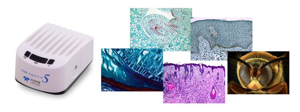
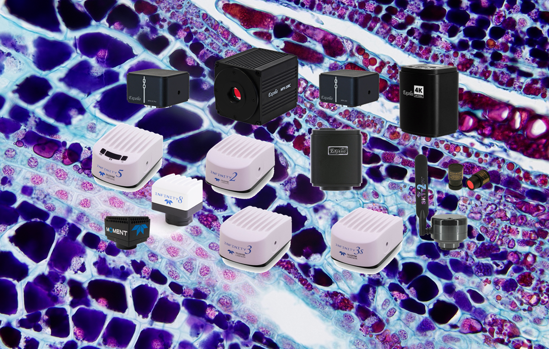
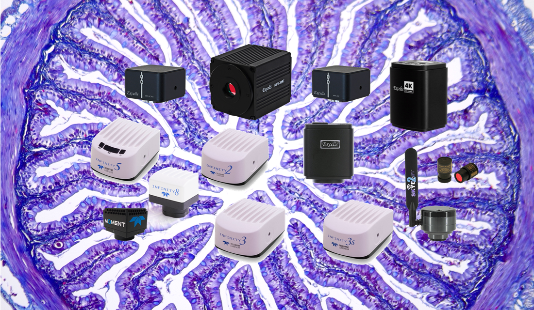
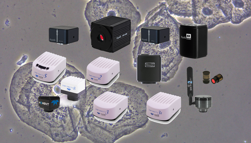
Now that you have a better understanding of the depth of focus vs. depth of field differences and how they affect the quality of microscopes, see their influences for yourself with the help of ACCU-SCOPE’s high-quality microscope products. We offer a range of upright, inverted and dissection microscopes and various microscope accessories for laboratory settings and other environments. To learn more information about our products, give us a call today.