How Does Fluorescence Microscopy Work?
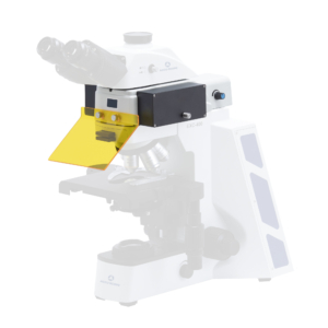
Preparing biological samples for fluorescence microscopy involves labeling them with fluorophores. These are fluorescent molecules that attach themselves to target elements within the sample, such as DNA or proteins. Once placed on the stage or inside the microscope, the sample is exposed to a specific wavelength of light generated by an LED, laser, or mercury lamp. The fluorophores absorb this light and release light at a longer wavelength and a different color. Using special optical emission filters and a dichroic mirror, the original emitted light is blocked and only the fluorescence emitted from the fluorophores in the sample can be seen through the eyepiece or camera. This produces a high-contrast image that enables the user to see the target elements with greater clarity than typical microscopy allows.