
Pathology Microscopes

Pathology microscopes provide the optical clarity and reliability laboratory professionals need to evaluate cells and tissues. Whether reviewing routine slides or documenting unusual findings, the right pathology microscope can support accurate observation, efficient workflows, and comfortable operation throughout the workday.
ACCU-SCOPE offers pathology microscopes designed to deliver the performance and precision clinical laboratories need at a strong overall value. With high-quality optics, dependable illumination, ergonomic configurations, and digital imaging options, ACCU-SCOPE systems support accurate specimen evaluation and efficient daily workflows without unnecessary complexity.

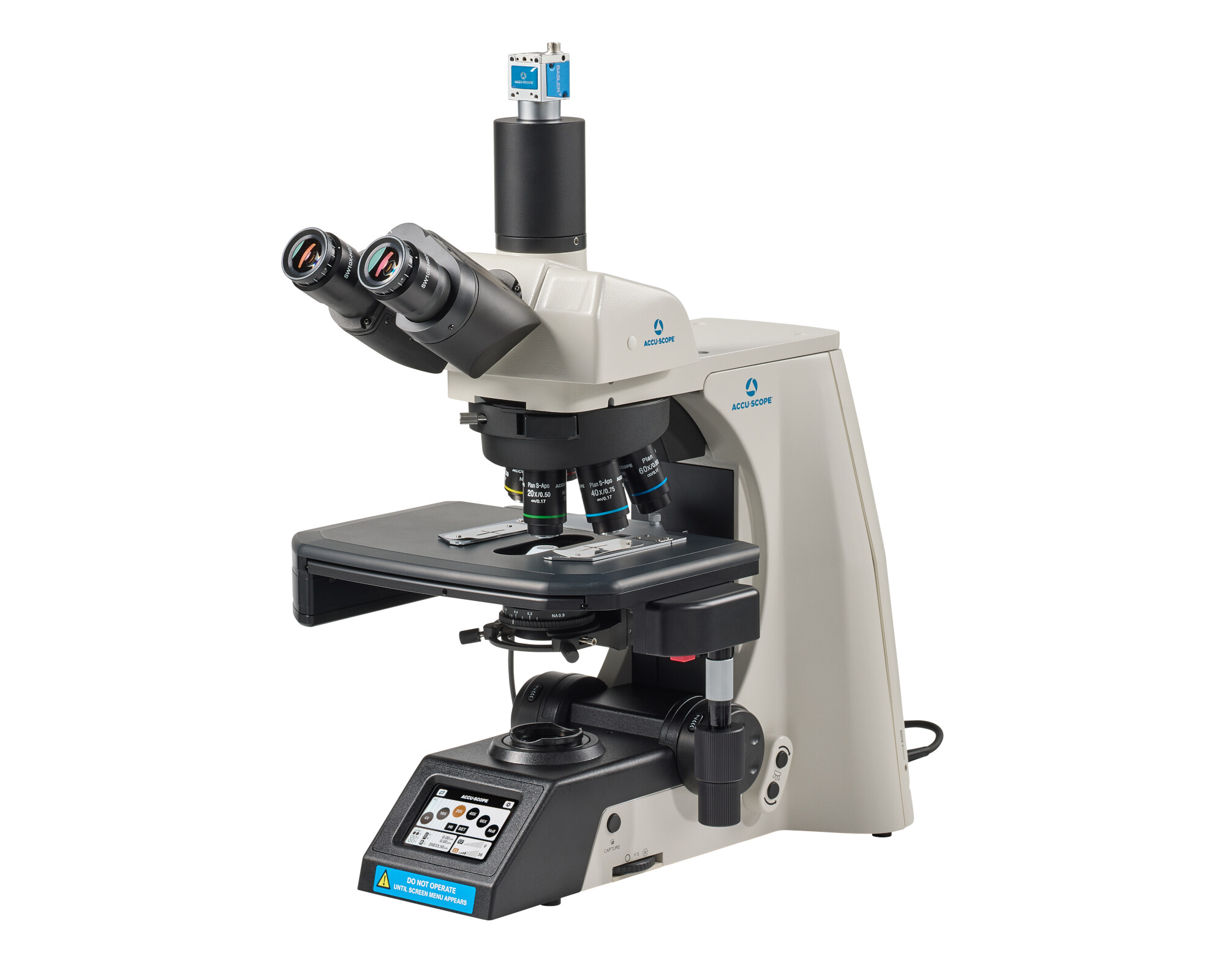
RC500

The ACCU-SCOPE RC500 remote collaboration system enables seamless automation and telepathology utilization, whether the user is sitting at the...
Learn More

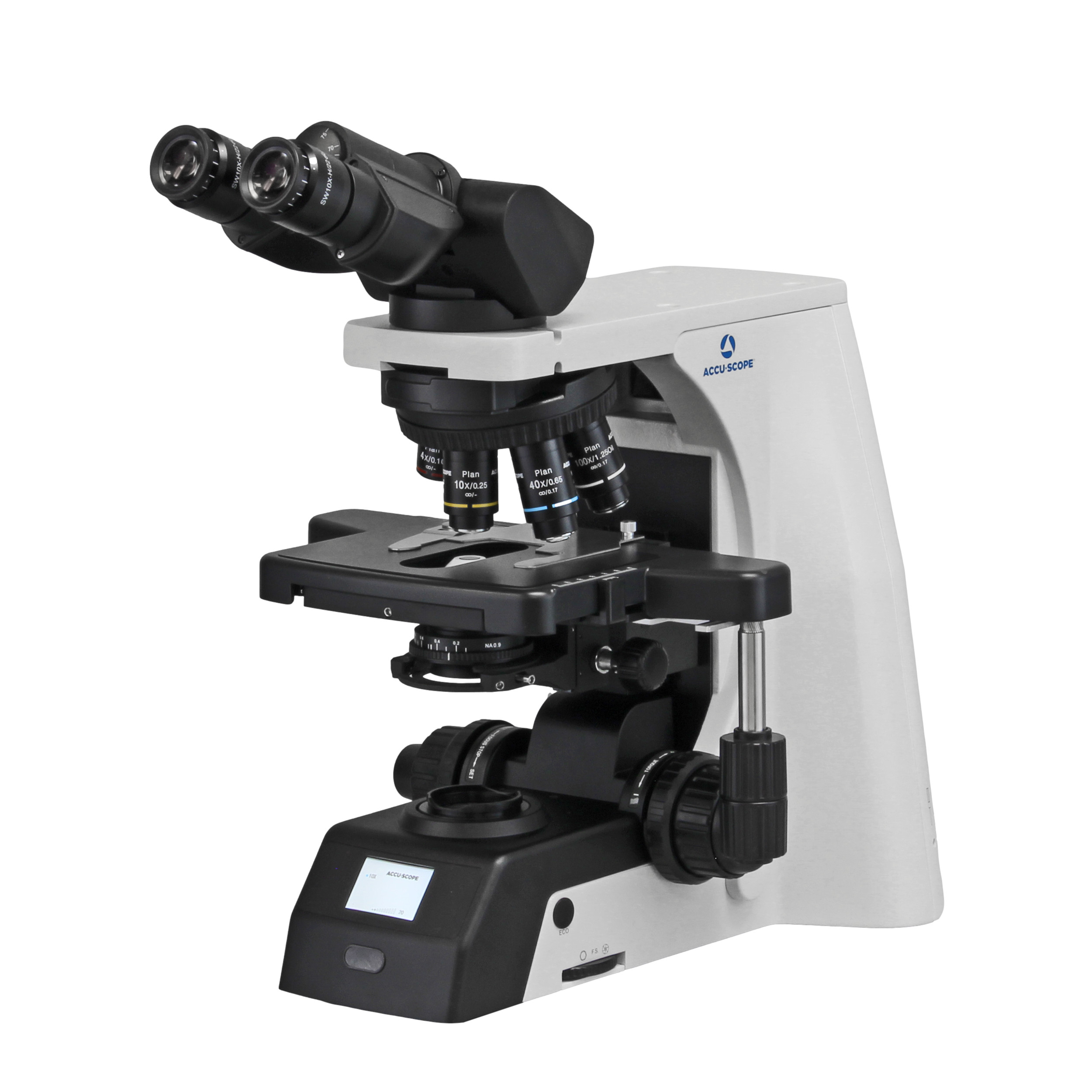
EXC-500

ACCU-SCOPE’s flagship EXC-500 Microscope offers best-in-class performance and value for clinical laboratory and research applications. The NIS...
Learn More

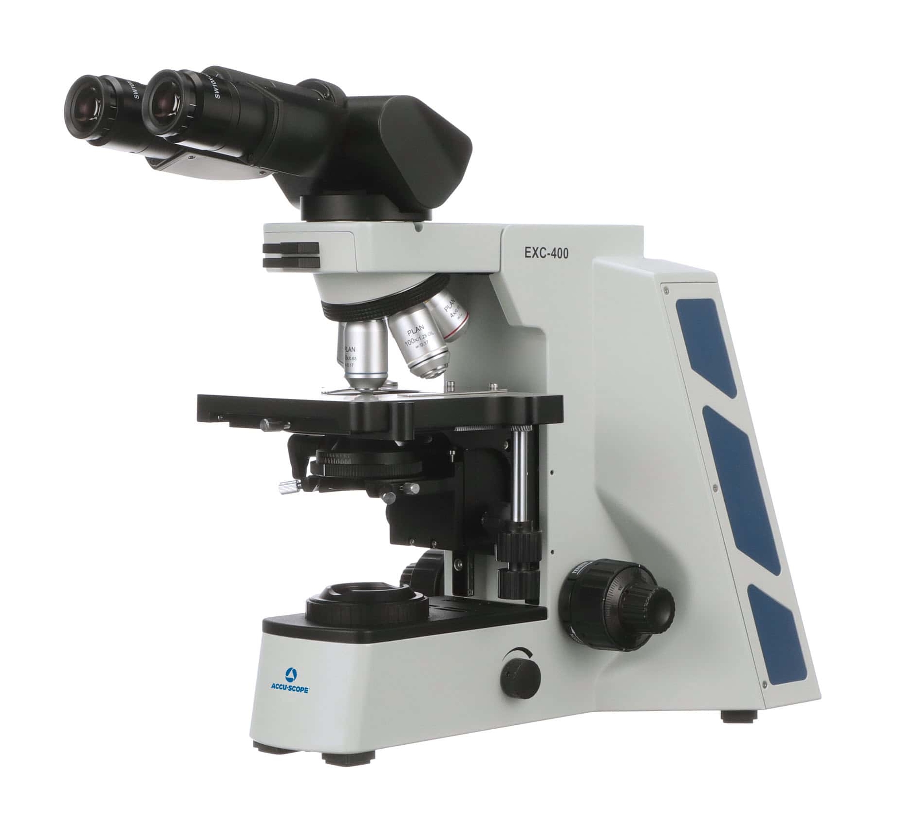
EXC-400

The EXC-400 from ACCU-SCOPE is designed for a broad range of microscopy applications in clinical, academic, and research environments. The EXC-400 is...
Learn More

What to Consider When Choosing Pathology Microscopes

Equipment Expectations for Clinical Laboratories

A microscope intended for routine clinical use needs to fit both the laboratory workflow and the physical workspace. Consider the available bench footprint, electrical connections, camera or computer placement, and access to existing laboratory information systems before choosing a configuration.

Laboratories should also verify that the microscope, camera, software, and related components meet the regulatory, validation, documentation, and intended-use requirements applicable to their facility. Service procedures, preventive maintenance, image calibration, and validation requirements should be established according to laboratory policies.

Read more

Optical Performance and Objective Choices

Objective selection has a direct effect on image quality. Plan Achromat objectives provide a flat field across the viewing area and are well suited to many routine clinical applications. Semi-apochromat and apochromat objectives provide progressively greater correction for chromatic and optical aberrations, making them valuable when demanding color reproduction, resolution, or digital imaging is required.

For pathology, low-power objectives are especially important because they allow rapid scanning of tissue sections before moving to higher magnification. A wide field of view can also reduce the amount of stage movement required when examining larger specimens.

Whatever objective class is selected, parfocal performance and consistent color correction help make transitions between magnifications faster and more comfortable.

Read more

HD Camera and Digital Imaging

A trinocular pathology microscope can be equipped with a digital camera for documentation, consultation, teaching, measurement, and image sharing.

Camera resolution should be selected according to the intended application rather than resolution alone. High-resolution cameras can preserve fine specimen detail, while a strong frame rate makes focusing, stage movement, and live viewing more responsive.

ACCU-SCOPE offers several digital imaging options. The Excelis 4K camera uses an 8.3 MP sensor, provides 3840 × 2160 resolution, and supports HDMI, USB 3.0, and Ethernet connections at up to 30 frames per second. CaptaVision+ software provides tools for measurement, annotation, image processing, reporting, and data export, with DICOM image support available in the Windows version.

For faster live imaging, the ALTRA PathoPix 4K provides 4K resolution with live output up to 60 fps through HDMI and USB, along with Ethernet and Wi-Fi connectivity options.

Read more

LED Illumination and Contrast

LED illumination offers several benefits for laboratories that operate microscopes for extended periods. LEDs provide consistent output, generate relatively little heat, and reduce the maintenance associated with frequently replacing traditional lamps.

Proper Köhler illumination helps deliver an evenly illuminated field with good contrast and resolution. Laboratories should periodically confirm illumination alignment and uniformity as part of routine microscope care.

When fluorescence is required, consider available excitation sources and filter configurations before selecting a microscope. The EXC-500 can be configured for fluorescence as well as brightfield, phase contrast, polarization, darkfield, and DIC observation.

Read more

Mechanical Design, Ergonomics, and Stage Options

Pathologists may spend hours each day at the microscope, making ergonomic design an important consideration. Adjustable viewing heads, interpupillary distance controls, accessible focus controls, and comfortable stage positioning can reduce unnecessary neck, shoulder, and arm movement.

The stage should provide smooth, precise movement for systematic examination of slides. Traditional mechanical XY stages offer direct manual control, while motorized stages can support automated scanning and remote workflows.

ACCU-SCOPE’s EXC-500 also offers a plain pathology stage without XY controls for users who prefer to manually position slides. Ergonomic binocular and trinocular viewing heads and eye-level risers are available for laboratories seeking additional operator adjustments.

Read more

Microscope Options for Pathology From ACCU-SCOPE

ACCU-SCOPE offers several upright microscope platforms appropriate for clinical laboratory and pathology applications. 

The EXC-500 is engineered for pathology and cytology applications and combines an infinity optical system with high-color-rendering LED illumination. Configurations include Plan Achromat, Plan Semi-Apochromatic, and Plan Apochromatic objectives, ergonomic viewing options, digital imaging, and multiple contrast methods.

The EXC-400 provides a durable upright platform with LED illumination, a rackless mechanical stage, and optional fluorescence, phase contrast, and darkfield capabilities. Its LED is rated for more than 20,000 hours of continued use.

For laboratories interested in remote consultation or live telepathology, the RC500 Remote Collaboration System combines a motorized stage, motorized objective selection, digital imaging, slide scanning, measurement, annotation, and remote microscope control. It is designed to support applications such as telepathology, consultation, frozen sections, FNA evaluation, education, and collaboration.

ACCU-SCOPE proudly serves the clinical sector with high-quality, high-performing microscopes and related accessories. Our microscopes and electronic components include a five-year limited warranty for the original end user, subject to the published terms. LED lamps carry one-year warranty coverage.

Frequently Asked Questions About Pathology Microscopes

What Magnifications Are Commonly Used in Pathology?

Pathologists commonly scan tissue at low magnifications (e.g., 2x or 4x) before moving to 10x, 20x, or 40x objectives for closer examination. The ideal configuration depends on the specimen and laboratory application.

Yes. Trinocular microscopes are designed to accommodate digital cameras. Confirm the camera mount, sensor size, optical adapter, software, operating system, and desired connectivity before configuring the system.

LED illumination generally provides a long operating life compared with conventional lamps. Individual ratings vary by microscope; for example, the EXC-500 LED is rated for more than 25,000 hours and the EXC-400 LED for more than 20,000 hours. Laboratories should still inspect illumination performance and follow the manufacturer’s recommended preventive maintenance procedures.