
Hematology Microscopes

ACCU-SCOPE hematology microscopes provide clinical laboratories, hospitals, veterinary practices, and teaching labs with the clarity needed to examine blood cells and bone marrow samples. Strong color fidelity, digital options, available Quick Ship configurations, and five-year limited microscope coverage combine professional performance with outstanding value.

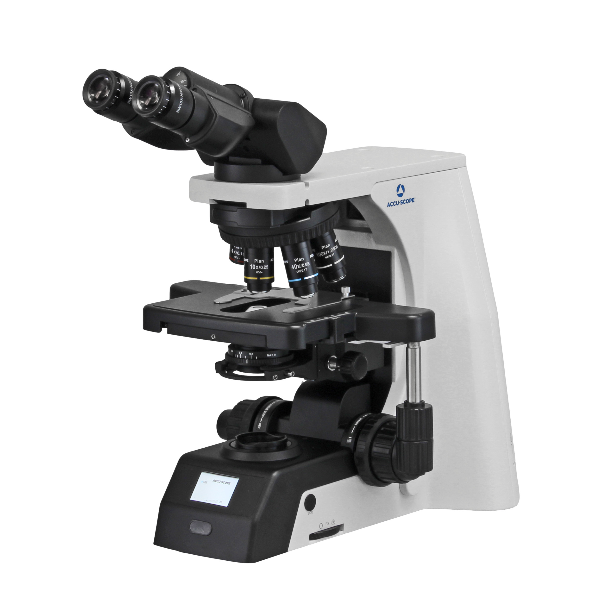
EXC-500

ACCU-SCOPE’s flagship EXC-500 Microscope offers best-in-class performance and value for clinical laboratory and research applications. The NIS...
Learn More

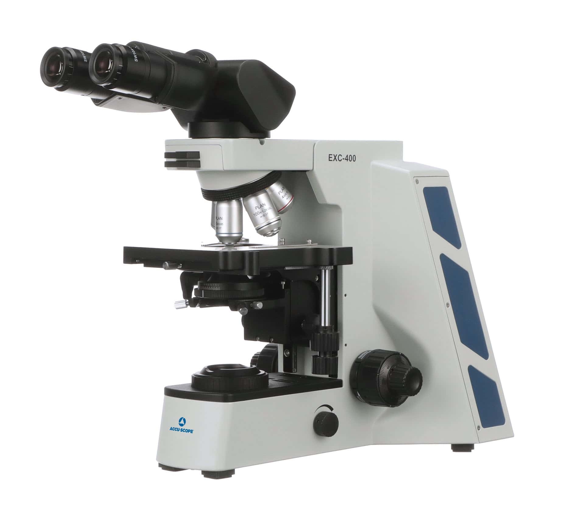
EXC-400

The EXC-400 from ACCU-SCOPE is designed for a broad range of microscopy applications in clinical, academic, and research environments. The EXC-400 is...
Learn More

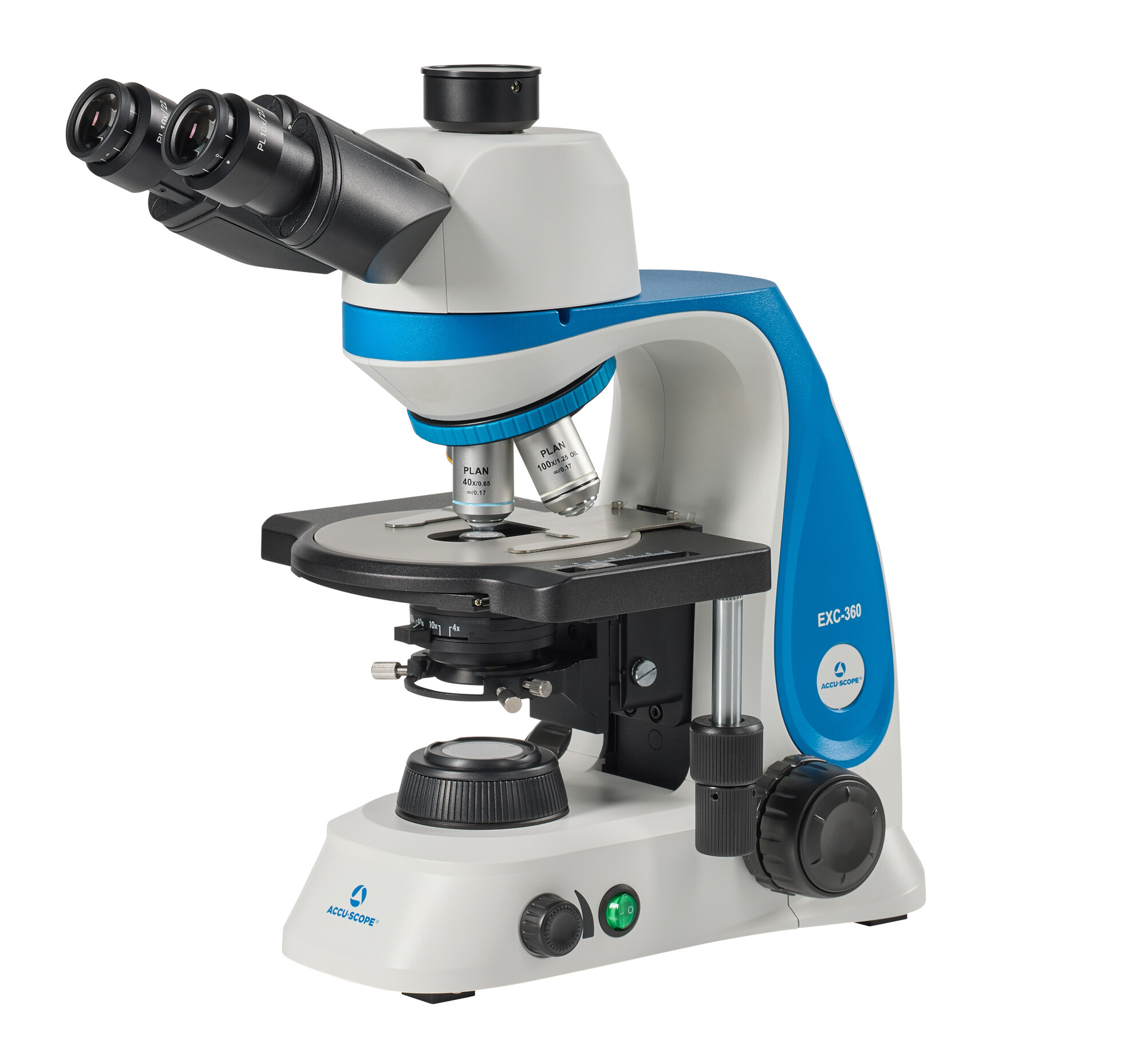
EXC-360

The NEW EXC-360 from ACCU-SCOPE delivers exceptional optical performance, versatility and durability for clinical, veterinary and education...
Learn More

Types of Microscopes for Hematology and Routine Blood Smears

Different hematology microscopes are designed for different purposes, such as supporting routine brightfield review, digital documentation, live-cell research, fluorescence assays, or sample preparation.

Upright Clinical Hematology Microscopes

Upright compound microscopes are the standard selection for a stained peripheral blood smear. Blood smears are typically prepared on glass microscope slides without a cover glass. Typical hematology configurations include 10x, 40x, 50x oil, and 100x oil objectives; with 10x eyepieces, the 100x objective provides 1000x total magnification for detailed review of red blood cells, white blood cells, platelets, and blood-borne parasites.

Read more

Digital Hematology Microscopes for Blood Cell Imaging

Digital hematology microscopes couple an optical system with a camera, monitor, or computer connection. A trinocular port or integrated camera allows a lab to archive representative fields, document white blood cell morphology, support quality review, and share images for consultation or telepathology.

USB cameras connect to image software, while HDMI or 4K systems can display live views without a computer and Wi-Fi models can send images to a phone or tablet. Browse ACCU-SCOPE digital imaging options before configuring a system, and always confirm the file formats, software compatibility, measurement tools, and storage policies ahead of ordering.

Read more

Inverted and Fluorescence Microscopes for Blood Cell Studies

Inverted microscopes are intended for blood cells growing in culture vessels, rather than routine slide review. Phase contrast can improve the visibility of transparent, unstained cells, while fluorescence reveals tagged targets in advanced cell research and selected hematology assays.

Read more

Stereo Microscopes for Sample Preparation and Veterinary Hematology

Stereo microscopes provide lower magnification and a greater working distance for sample handling, dissection, sorting, and preparation. They aren’t the primary choice for a stained blood film, but they can help veterinary teams with specimen preparation.

Read more
Feature image

Why Choose ACCU-SCOPE?

ACCU-SCOPE builds hematology microscopes for precision, performance, and value. Our U.S.-owned and operated facilities streamline assembly, design, quality control, sales, and technical support. Products are tested and inspected under documented quality processes, with select systems available through Quick Ship.

ACCU-SCOPE microscopes and electronic components carry a five-year limited warranty from the invoice date for the original end user, subject to the written terms. LED lamps carry one-year coverage. Ask our team for details on eligible models, bulk pricing, application support, and current delivery times.

What to Look for in Microscopes for Hematology

Optics and Objectives

Plan Achromat or Infinity Plan optics provide a flat, sharp field that’s great for comparing cell size, shape, and detail. Routine hematology microscopes offer 10x and 40x dry objectives plus 50x and 100x oil options for fine morphology or parasites.

Illumination and Contrast Methods

Köhler illumination balances brightness and contrast. A color-balanced LED can preserve subtle differences in Wright- or Giemsa-stained blood cells. Brightfield suits stained slides, while phase contrast is best for unstained, living specimens. Fluorescence requires the right filters, illumination, and labeled targets.

Stage Ergonomics and Workflow

A precise mechanical or rackless XY stage allows repeatable scanning. Low-positioned controls, an ergonomic head, and accessible focus knobs all reduce awkward positioning during long sessions; heavy-use labs should look for a rigid stand and metal gears.

Digital Imaging and Connectivity

Select a trinocular head or built-in camera for documentation, teaching, remote consultation, or quality review. Match the resolution, frame rate, field of view, interface, software, and file type to the task; the camera should faithfully reproduce stained blood cells.

Durability, Service, and Warranty

Compare warranty exclusions, service access, parts availability, and turnaround times. ACCU-SCOPE provides U.S.-based support, a service-center network, and Quick Ship for selected configurations. Read the warranty terms for full details.

How to Choose the Right Hematology Microscope for Blood Smears

First, focus on the specimen and daily workload. Routine blood smear review calls for an upright brightfield microscope with Plan optics, a precise stage, 10x and 40x objectives, and an oil-immersion objective for high-detail work. Choose a trinocular configuration when imaging, training, or telepathology are in the workflow.

For cultured blood cells, fluorescence, or time-lapse imaging, consider an inverted research system. Compare optics and support, then request a custom quote.

Frequently Asked Questions About Hematology Microscopes

What microscope can see blood cells?

A compound brightfield microscope shows stained red blood cells, white blood cells, and platelets. Clinical hematology microscopes commonly have 10x eyepieces and objectives from 10x through 100x; the 100x oil objective provides the resolution for detailed morphology and parasite review.

You can see the cell color, central pallor, and patterns associated with some hemoglobin-related blood disorders. However, individual hemoglobin molecules are below a standard light microscope’s resolution, so laboratories use hematology analyzers or other assays.

Hematology laboratories count on cell counters, slide makers and stainers, centrifuges, coagulation analyzers, flow cytometers, and microscopes. Automated instruments count and classify cells, while hematology microscopes let professionals review a peripheral blood smear, verify results, examine white blood cell morphology, and recognize sickle cells or schistocytes. Bone marrow evaluation relies on microscopic review, too.

An upright brightfield model with Plan optics, a mechanical stage, and dry and oil objectives is a good fit for most routine work. When the lab captures and shares images, choose a trinocular model.

Oil immersion supports detailed cell morphology and parasite examination at 1000x total magnification; lower powers help check smear quality and locate the monolayer.

Cameras help labs with documenting findings, teaching, consulting remotely, and retaining images for quality review. Accurate results depend on exposure, white balance, calibration, file management, and optical compatibility.

Keep the lenses, eyepieces, condenser, and stage clean; remove immersion oil promptly, and only with approved materials. Schedule regular checks for illumination, stage movement, focus, and camera calibration, following the manual and laboratory quality-control plan.