
Tissue Culture Microscopes

Tissue culture microscopes help researchers examine living cells, assess growth, and check the condition of cultures during daily laboratory work. Inverted microscopes are especially useful for cell culture because their objectives sit beneath the specimen, allowing users to view cells through the bottom of a culture vessel.

ACCU-SCOPE offers research microscopes and digital imaging solutions for university, biotechnology, and life science laboratories. Microscope selection depends on the samples being studied and the required imaging capabilities.

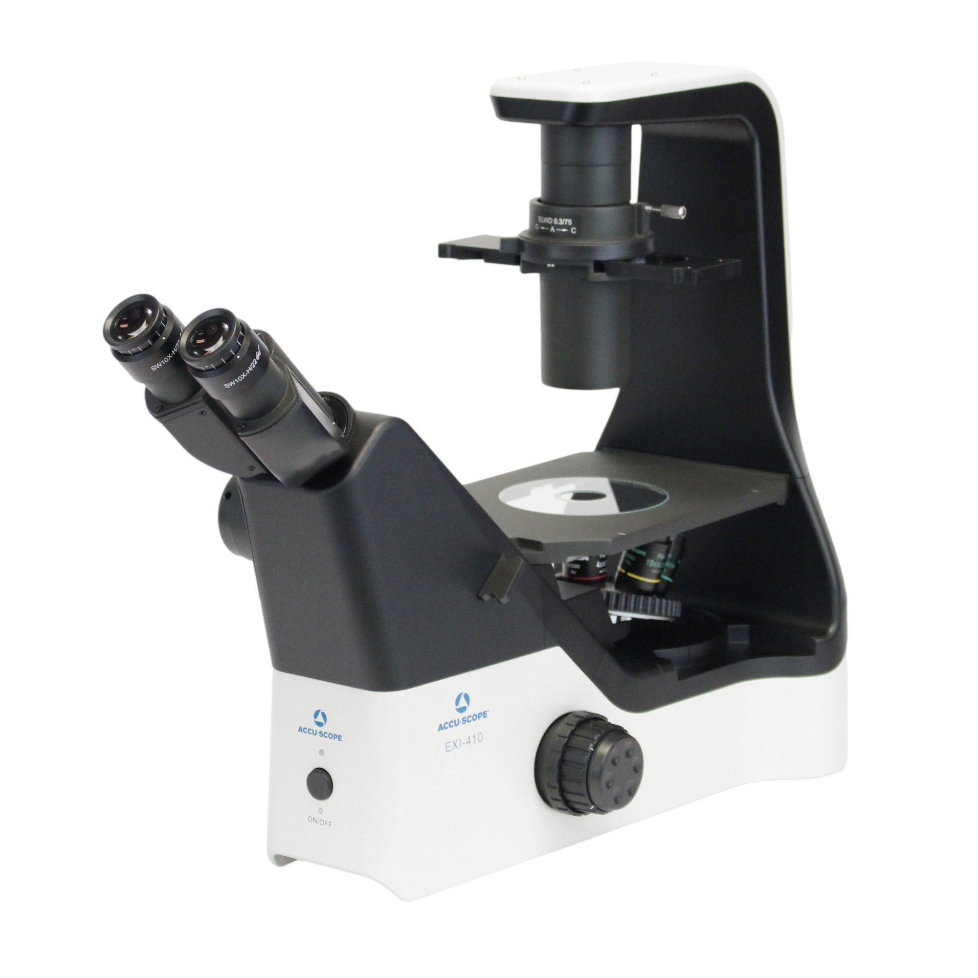
EXI-410

ACCU-SCOPE’s EXI-410 Inverted Microscope delivers outstanding optical performance, convenience, flexibility and value. Ideal for the daily...
Learn More

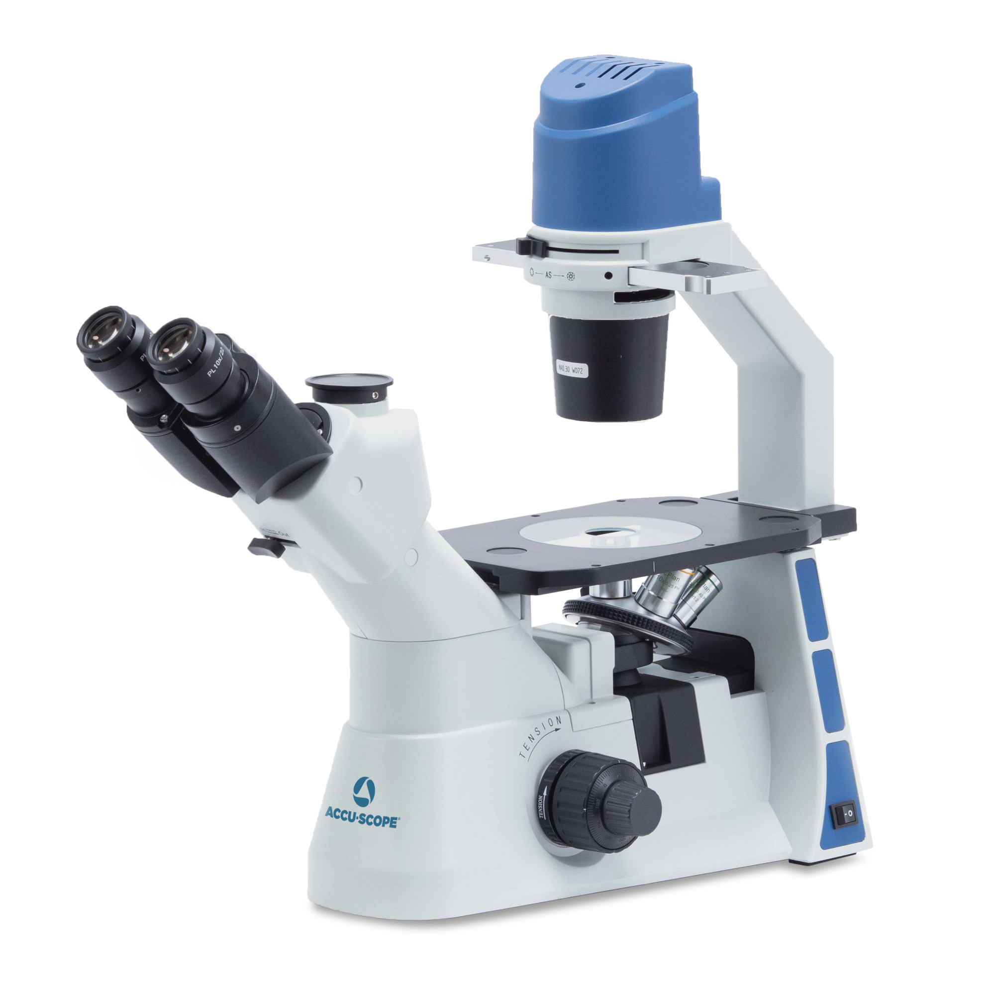
EXI-310

Utilizing ACCU-SCOPE’s AIS optical system and LED illumination for both diascopic (transmitted) and epi-fluorescence imaging, the EXI-310...
Learn More

What Is a Tissue Culture Microscope?

A tissue culture microscope is used to observe living cells and tissues as they grow in laboratory culture. Researchers use these instruments to inspect cell shape, growth patterns, and confluency, or the proportion of a culture surface covered by cells. Visible changes may suggest culture stress or contamination, although microscopy can’t detect every contaminant.

In an inverted microscope, the objectives are located below the stage and light is typically directed through the specimen from above. Cells growing on the bottom of dishes, flasks, and multiwell plates can be examined directly in their vessels. Upright microscopes commonly examine slides; stereomicroscopes suit larger specimens.

Types of Inverted Microscopes for Tissue Culture

Inverted microscopes are available in configurations for routine culture inspection, fluorescence microscopy, and advanced imaging applications.

Routine Inverted Microscopes

Routine models support checks of cell growth, shape, and culture condition. Brightfield imaging works well for samples with sufficient natural contrast or staining. Phase contrast microscopy makes transparent, unstained adherent cells easier to see, so it’s widely used during routine cell culture observation.

Fluorescence Inverted Microscopes

Fluorescence microscopy uses labeled cells or cellular structures to reveal specific targets. Depending on the system, illumination may come from LEDs or mercury-based light sources. Fluorescence can be useful in drug discovery, toxicology, cancer research, and regenerative medicine, but it’s not required for ordinary culture inspection.

Digital Inverted Microscopes

An inverted microscope equipped with a compatible digital camera can record images for reference, sharing, and comparison. Imaging software may assist with capture, organization, and measurements. Advanced analysis and automation depend on the configuration.

Essential Features When Selecting a Cell Culture Microscope

Start with the culture vessel, the detail you need to observe, and the frequency of use.  

Contrast Methods and Illumination

Brightfield is useful for many general observations, while phase contrast makes low-contrast living cells more visible without stains or fluorescent labels. Fluorescence is appropriate when a study calls for labeled structures or specific cellular signals, or the cells are expressing fluorescent proteins such as Green or Red Fluorescent Protein (GFP, RFP, respectively). Stable LED illumination supports routine viewing and generally places less heat near the specimen than some traditional light sources. Limit light exposure for sensitive live-cell studies.

Objectives, Working Distance, and Vessel Compatibility

Lower magnification provides a broader view for checking a culture; higher magnification reveals more cellular detail. Long working distance objectives can provide the clearance needed to focus through thicker culture vessel bottoms. Check optics and stage compatibility with dishes, flasks, and multiwell plates.

Stable stages and vessel holders support repeated observations. For some researchers a plain flat stage is sufficient to hold the tissue culture vessels for manual positioning, while other researchers may require a mechanical stage for more precise positioning such as when observing cultures in multiwell plates.  Consider camera ports and accessory options for future work.

How to Select the Right Tissue Culture Microscope

Begin by identifying the specimen and culture vessel. For routine observation of unstained adherent cells, an inverted microscope with phase contrast is often a practical starting point. Next, consider magnification, working distance, vessel holders, and the need for stable, repeatable positioning.

For documentation or more detailed research, look for a camera-compatible configuration with software suited to the planned analysis. Future fluorescence and automation requirements should also factor into equipment selection.

Tissue Culture Microscope Applications in Research

Research goals shape the optical and imaging requirements of cell culture microscopes.

Drug Discovery and Toxicology

Researchers observe how cultured cells respond to experimental compounds, including visible changes in shape, growth, or survival. Inverted microscope imaging can support repeatable observation across multiple wells.

Read more

Regenerative Medicine and Cell Research

Stem cell and regenerative medicine projects often require observation of cell growth, migration and changes in appearance during differentiation. Tissue culture microscopes also support cancer research and other cell-based studies where viewing live cultures with limited disturbance is valuable.

Read more

Clinical Research and Education

Clinical research laboratories may use inverted microscopes to examine experimental cultures. Academic laboratories also use them to teach students about cell growth, microscopy techniques, and routine culture assessment.

Read more

Digital Imaging and Analysis for Live Cell Research

Digital imaging creates a record of cell morphology and growth that researchers can revisit or share with colleagues. Camera selection depends on the required detail and observation method. Fluorescence work may demand greater sensitivity under low light, while resolution, frame rate, and software support also affect image capture.

Image Analysis and Documentation

With suitable software, image analysis can measure features such as cell count, cell shape or the area covered by a culture (confluency). Saved images also provide a reference for later review. Automated acquisition may reduce operator-to-operator variation when the imaging system and analysis process are properly controlled.

Environmental Control for Extended Imaging

Live cell imaging across longer periods requires attention to temperature, humidity, and the culture environment. Depending on the experiment, a compatible stage-top chamber or incubator arrangement may be needed. Repeated image capture allows researchers to document changes in cell behavior while limiting disturbance to the culture. Check accessory compatibility before selecting a system.

Frequently Asked Questions About Tissue Culture Microscopes

Why Is an Inverted Microscope Used for Tissue Culture?

Its objectives sit below the culture vessel, allowing researchers to observe cells growing on the bottom without transferring them to slides.

The best microscope depends on the cells, vessel type, and imaging tasks. Routine inverted microscopes with phase contrast suit many general cell culture applications. More specialized work may call for fluorescence, digital imaging, or environmental accessories.

Phase contrast microscopy increases the visibility of transparent, unstained cells. It helps users observe cell shape, growth patterns, and culture condition during routine checks.

Yes. Compatible cameras can capture images of living cells, and suitable software may support repeated acquisition such as time-lapse or video capture. Longer experiments may also require environmental controls.

Explore ACCU-SCOPE Tissue Culture Microscopes

Compare ACCU-SCOPE inverted microscope and digital imaging options with your laboratory requirements in mind. Consider contrast methods, vessel compatibility, objectives, and documentation needs. Contact our team for application guidance or connect with an authorized distributor to discuss the appropriate configuration for your research.